
Case Presentation
Austin J Radiol 2023; 10(1): 1209
Intra-Peritoneal Rupture of a Benign Ovarian Teratoma - A Rare Complication of This Relatively Common Entity
Arora R1*, Desai P2, Jikadara P3 and Deshpande S4
1Assistant Professor, Dept. of Radiodiagnosis and Imaging, Government Medical College and New Civil Hospital, Surat
2Professor and Head, Dept. of Radiodiagnosis and Imaging, Government Medical College and New Civil Hospital, Surat
3Senior Resident, Dept. of Radiodiagnosis and Imaging, Government Medical College and New Civil Hospital, Surat
4Senior Resident, Dept. of Radiodiagnosis and Imaging, Government Medical College and New Civil Hospital, Surat
*Corresponding author: Rajat Arora Department of Radio diagnosis and Imaging, Government Medical College and New Civil Hospital, Surat, Bungalow no. 2, behind Kapadia Health Club, Janta Nagar-B, New Civil Road, Surat, Gujarat, India.
Received: November 30, 2022; Accepted: January 03, 2022; Published: January 09, 2023
Abstract
Mature cystic teratoma is a cystic tumor composed of welldifferentiated derivations from at least two of the three germ cell layers (ectoderm, mesoderm, and endoderm). These tumors are usually complicated by torsion, with rupture and malignant transformation to be relatively rare. Here we present a case of a 25-year-old woman with complaints of sudden onset generalized acute abdominal pain. Her ultrasound pelvis revealed a large anechoic lesion with internal chunky calcification as well as hyperechoic internal contents (like that of fat) in the pelvis and left adnexa, extending up to the midline. Mild to moderate loculated ascites, peri-hepatic and intraperitoneal nodularity were also seen, representing changes of peritonitis.
On CECT abdomen and pelvis, focal discontinuity was seen within the pelvic cyst wall with evidence globular free fat contents within the peritoneal cavity. Findings related to peritonitis were corroborated on CT.
The imaging findings were suggestive of underlying peritonitis, secondary to the intra-peritoneal rupture of dermoid and release of its internal fat and fluid contents.
Keywords: Dermoid; Teratoma; Intraperitoneal; Rupture; Ultrasound; Peritonitis
Case presentation
A 25 years old multiparous female presented with complaints of sudden onset generalized acute abdominal pain, with h/o intermittent pelvic pain was present. However, her menstrual cycles were regular.
Her ultrasound pelvis revealed a large anechoic lesion with internal chunky calcification as well as hyper-echoic internal contents (like that of fat), seen in the pelvis, in the left adnexa, extending up to the midline (Figure 1). Apart of this large lesion, mild to moderate loculated ascites was seen (Figure 2). Peri-hepatic and intraperitoneal nodularities were seen, which represented changes of peritonitis (Figure 3).
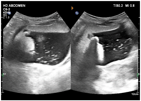
    Figure 1: USG image showing a well-defined large anechoic lesion

with internal calcification chunks (with post acoustic shadowing)

and hyper echoic internal contents (like that of fat).
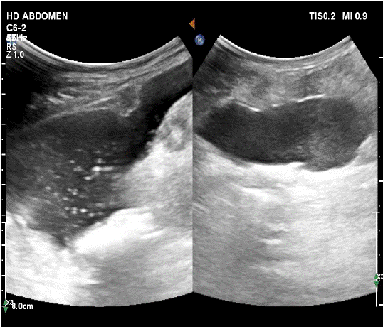
    Figure 2: Image on the right shows loculated ascites with echoes

and peritoneal nodularity Image on the left shows an anechoic intraperitoneal

tubular lesion, with its appearance seen distinct from

the ascitic fluid.
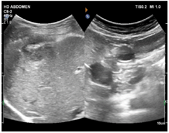
    Figure 3: Image on the right shows perihepatic nodularity seen, indenting

the hepatic surface. Image on the left shows an anechoic

lesion, seen in the peripancreatic region.
However, the underlying cause of her peritonitis couldn’t be justified. So she was advised CECT abdomen and pelvis study for evaluation of the underlying cause of peritonitis. CT showed the teratoma, with its calcific and fat contents (Figure 5) with globular free fat contents within the perihepatic nodules as well as within the peritoneal cavity (Figure 4 and 7). A large fat containing tract was seen, extending from the large pelvic lesion into the peritoneal cavity on the right side, with focal discontinuity seen within the cyst wall (Figure 6). A chunk of calcification was also seen lying freely in the peritoneal cavity, outside the pelvic lesion.
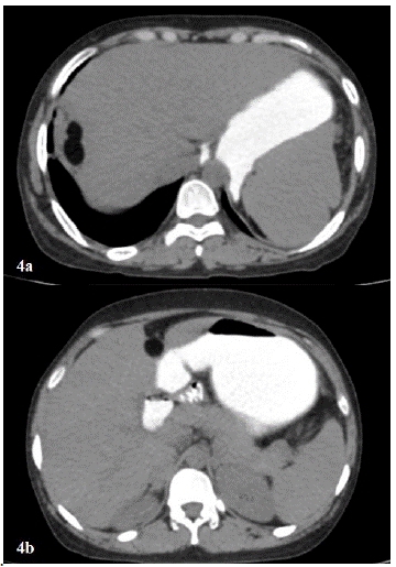
    Figure 4: Plain CT axial image of upper abdomen shows (a) perihepatic

nodule with fat content in it and (b) another small fat globular

nodule in the peripancreatic region, Corroborating with the USG

images.
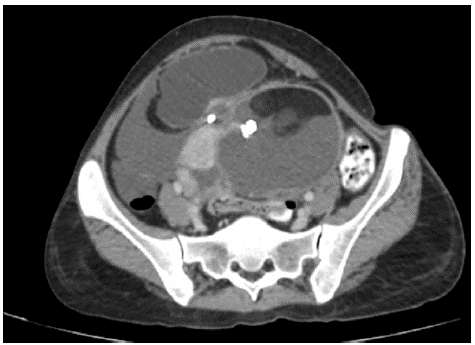
    Figure 5: CECT axial image of pelvis shows typical fat and calcification

containing lesion in the left adnexa - Typical for a mature cystic

teratoma.
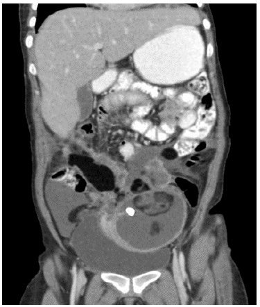
    Figure 6: CECT coronal image, showing dermoid, the fat containing

track in the peritoneal cavity extending from the teratoma (marked

by blue arrow) as well as loculated ascites.
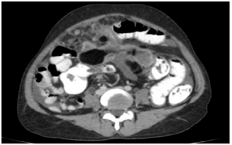
    Figure 7: CECT axial image of pelvis shows peritoneal nodularity

and omental caking. The fat containing track (mentioned above)

also seen in this axial image.
Ascites and enhancing peritoneal-perihepatic nodularities were suggestive of underlying peritonitis, secondary to the intra- peritoneal rupture of dermoid and release of its internal fat and fluid contents.
Based on these imaging findings, diagnosis of benign ovarian teratoma, with its intra-peritoneal rupture and resultant secondary inflammatory changes of peritonitis was made. The other differentials for the imaging findings included pseudomyxoma peritonei, tuberculous peritonitis, and primary peritoneal neoplasm with co-existing dermoid and malignant transformation of invasive teratoma with peritoneal metastases
The radiological findings were corroborated intra-operatively and on Histopathology.
Intra-operatively, extensive bowel and peritoneal adhesions, peritoneal nodules and fat globules were found. Limited partial excision of lesion was possible due to adhesions. Histopathology from the excised lesion and omentum revealed features of benign well-differentiated teratoma and chronic granulomatous changes respectively.
Final diagnosis was benign ovarian teratoma with its intraperitoneal rupture and secondary chronic peritonitis.
Discussion
Mature cystic teratoma (or more commonly “dermoid cyst”) is a cystic tumor composed of well-differentiated derivations from at least two of the three germ cell layers (ectoderm, mesoderm, and endoderm). They affect a younger age group (mean patient age, 30 years) than epithelial ovarian neoplasm [1].
Mature cystic teratoma is the most common germ cell neoplasm [2,3]. It is the most common ovarian mass in children.
On imaging, most of these lesions can be easily diagnosed on USG. Multiple appearances are well-known for these lesions i.e., echogenic bands, fat-fluid levels, echogenic nodules (dermoid plug, dermoid mesh), dot and dash appearance due to variable composition of keratin, sebum, fat, hair and calcifications.
However, these lesions have certain complications including torsion, rupture, malignant transformation, infection and autoimmune hemolytic anemia.
Reported incidences of these complications show torsion to be the most common and rupture and malignant transformation to be relatively rare.
Management of each of these complications is also very different. Therefore, timely and accurate diagnosis of these complications is important for optimal patient treatment.
In our patient, we saw intra-peritoneal rupture of dermoid, which caused leakage of the liquefied sebaceous contents, fat as well as calcific chunks into the peritoneum, which irritates the peritoneum and leads to acute or chronic inflammation.
Although the prognosis of chronic rupture is favorable, dense peritoneal adhesions caused by chronic recurrent peritonitis may cause other secondary complications such as bowel obstruction.
In our patient, we also suspected that underlying torsion could have been the inciting event for rupture of teratoma, as the cyst was reaching upto the midline.
Written informed consent of the patient for publication was obtained.
Conclusion
In a patient with teratoma with complaints of abdominal pain, secondary complications associated with teratoma should be evaluated, to rule out torsion and its rupture.
Majority cases of teratoma can be easily diagnosed with USG.
CT is very helpful while assessing the secondary complications of teratoma.
References
- Omerci JT, Licciardi F, Bergh PA, Gregori C, Breen JL. Mature cystic teratoma: a clinicopathologic evaluation of 517 cases and review of the literature. Obstet Gynecol. 1994; 84: 22-28.
- Koonings PP, Campbell K, Mishell DR, Grimes DA. Relative frequency of primary ovarian neoplasms: a 10-year review. Obstet Gynecol. 1989; 74: 921-926.
- Whitecar MP, Turner S, Higby MK. Adnexal masses in pregnancy: a review of 130 cases undergoing surgical management. Am J Obstet Gynecol. 1999; 181: 19-24.
- Pantoja E, Noy MA, Axtmayer RW, Colon FE, Pelegrina I. Ovarian dermoids and their complications: comprehensive historical review. Obstet Gynecol Surg. 1975; 30: 1-20.