
Case Report
Austin J Radiol. 2023; 10(1): 1210.
Help, My Lung is Trapped! – A Case Report
Naura A, Neupane NP*, Regmi A and Rajlawot K
Radiologist, Shahid Gangalal National Heart Centre, Bansbari, Kathmandu, Nepal
*Corresponding author: Nirmal Prasad NeupaneRadiologist, Shahid Gangalal National Heart Centre, Bansbari, Kathmandu, Nepal
Received: December 06, 2022; Accepted: January 24, 2022; Published: January 31, 2023
Abstract
Trapped lung is defined as the inability of the lung to expand fully and fill up the thoracic cavity because of a restricting fibrous visceral pleural layer preventing apposition of visceral and parietal pleura. It is a sequela of a previous inflammation of the pleural space and has variety of causes. Chest radiograph and thoracic ultrasound is the key imaging modalities for the diagnosis usually with a history of preceding pleural drainage. Typical imaging features include visceral pleural peel, basal pneumothoraces, ipsilateral volume loss and lobar atelectasis. Management aims at relief of symptoms and enabling full lung re-expansion. It is usually associated with high morbidity, therefore identifying patients with trapped lung is crucial for early treatment and positive patient outcomes. We present a case of trapped lung of a patient with previous history of pulmonary tuberculosis and chronic right pneumothorax.
Introduction
Trapped lung is defined as the inability of the lung to expand fully and fill up the thoracic cavity because of a restricting fibrous visceral pleural layer (peel) that prevents normal visceral and parietal pleural apposition [1]. It occurs as a result of a remote pleural space inflammation or a long-standing stable pleuro-pulmonary disease, either from an inflamed lung or a malignant visceral pleural tumor. This results in formation of a mature, fibrous visceral pleural membrane that prevents the lung from re-expanding, leading to a negative pressure environment in the pleural space. Negative pleural pressure within the pleural space increases the entry of fluid into pleural space and reduces pleural fluid exit through pleural lymphatics thus leading to the formation of a chronic pleural effusion. When the pleural fluid is removed during thoracocenteses in these cases, the fibrotic restrictive visceral pleura prevents the lung from expanding, leaving air trapped between parietal and visceral pleura, radio graphically identified as a pneumothorax ex vacuo [2-4].
To identify and treat the underlying causes of pleural inflammation and to avoid the progression to trapped lung and high morbidity associated with multiple invasive diagnostic procedures, early evaluation of pleural effusion is very important, although pleural fluid drainage might not always provide clinical relief to the patients [1,5]. Herein, we describe a case of trapped lung who presented to our hospital with chronic unilateral pneumothorax.
Clinical History
A 62-year-old woman referred to the Emergency Department (ER) of our hospital from medical outpatient department, for chest tube insertion for right sided pneumothorax that was detected on chest X-ray. She presented to the hospital with the complaint of sudden onset of shortness of breath since last 5 hours. She had reportedly experienced few similar occurrences in last 9 months. She is a known case of Diabetes Mellitus (DM) and Hypertension, on medications (Furosemide 40mg, Ramipril 50mg, Empagliflozin 10 mg and subcutaneous injection insulin glargine). She denied any preceding history of cough, sputum production, chest pain, fever, or wheezing. She was diagnosed to have Pulmonary Tuberculosis (PTB) in her childhood, when she as 12 years of age, and had underwent antitubercular therapy then. She also had given history of right sided pneumothorax that had occurred about 10 years ago, for which she did not receive any treatment as the pneumothorax was minimal and she was asymptomatic. Moreover, she never had any follow-up or review done for the right sided pneumothorax. In ER, the patient was a febrile with a temperature of 36.7°C, heart rate was 98 beats per minute, blood pressure was 105/72 mmHg, respiratory rate was 24 breaths per minute and SpO2 was 88% on room air. Current physical examination revealed an absence of breath sounds on the right side but no dullness on percussion. The rest of the examination was unremarkable. Chest X-ray (done before coming to ER) was consistent with right sided pneumothorax with meniscus sign in right costo-phrenic angle indicating that this is actually a hydropneumothorax. Due to this, further inquiring was done and patient revealed that a right pleural effusion was once treated with pleural catheter placement, about 6 months ago in their regional hospital (no medical records available). In our ER, she was finally advised for Computed Tomography (CT) scan of Chest. For the publication of the case contents and images, informed written consent was taken from the patient prior to data collection.
Imaging Findings
The CT images of the patient showed a large right sided pneumothorax (Figure 1) with abnormal thickening of visceral pleura (Figure 2), thick adhesive bands (Figure 3) and minimal right pleural effusion resulting in right sided hydropneumothorax (Figure 4). In addition to this, partial collapse of right lung with fibrobronchiectatic and fibrocalcific changes were noted (Figure 2 and 3). In the left lung, mosaic attenuation pattern (Figure 1) was also noted indicative of air-trapping in addition to few fibrotic changes (Figure 4). The aforementioned findings are in consistent with Trapped lung.
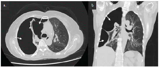
Figure 1: Right sided pneumothorax. An axial (a) and coronal (b) CT lung window images showing a large air-attenuating area in right pleural space (arrows).
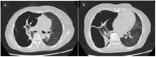
Figure 2: CT axial lung window images (a) and (b) showing the thickened visceral pleura giving rise to “fibroticpleural peel” that prevented the lung from re-expanding (arrows). Partial collapse of right lung can be appreciated (b).
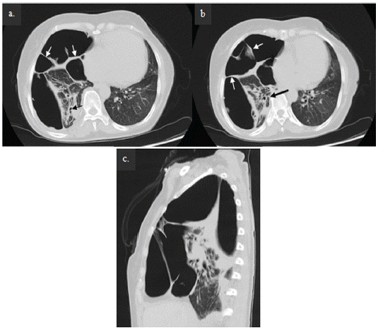
Figure 3: CT axial images (a) and (b) and sagittal image (c) of lung window showing the thick adhesive bands between the thickened visceral pleura and normal parietal pleura (white arrows). Fibrobronchiectatic and fibrotic changes (long black arrow) can also be appreciated in the partially collapsed right lung.
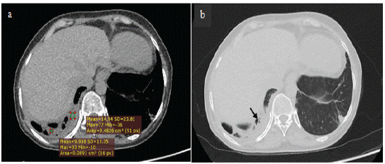
Figure 4: Plain CT axial image (a) and lung window axial image (b) of basal lung showing minimal pleural effusion (HU +10 to +15) with few small air locules in the basal region (arrow) suggestive of hydropneumothorax. Fibrotic changes in basal left lung can also be appreciated.
Discussion
Trapped lung is characterized by the atelectatic lung that is unable to expand and fill the thoracic cavity, due to a restricting fibrous visceral pleural peel. To diagnose trapped lung, drainage of the pleural space must result in both radiographic findings of a resultant pneumothorax ex-vacuo and a thickened visceral pleura [6]. The resulting fluid filled chronic pleural space, and the persistence of the fluid is solely due to hydrostatic equilibrium [7]. If pleural manometry is performed, negative intrathoracic pressure is identified and elevated pleural elastance can be calculated. Trapped lung, being a sequela of a previous inflammation of the pleural space, can have various causes. It was historically recognized as a complication of therapeutic pneumothorax for treatment of tuberculosis. More recent studies show it to be a consequence of inadequately treated parapneumonic effusion, commonly associated with cardiac surgery, chest trauma, and other inflammatory processes involving the pleura such as malignant pleural effusion [6,7]. The diagnosis of trapped lung requires chronicity and stability and the absence of active inflammatory or malignant pleural process, bronchial obstruction or any severe underlying lung disease [7]. Considering the history of chronic pneumothorax and history of thoracocentesis with persisting post-thoracentesis pneumothorax, clinical examination including chest X-ray and CT scan findings and the absence of other possible etiologies, our clinical diagnosis was trapped lung caused by remote pneumothorax, all of which possibly occurring secondary to pulmonary tuberculosis several years ago. There are various treatment options for trapped lung, and the choice of treatment modality is determined by the patient's symptoms and pathology. If the effusion is not worsening and the patient is asymptomatic, observation is warranted. A more definitive approach, surgical decortication is indicated for symptomatic patients, which is the only curative treatment modality [1,8]. In our case, due to incapacitating dyspnea, pleural decortication was advised and patient was referred to Department of thoracic surgery of another dedicated thoracic centre for definitive treatment.
Conclusion
Trapped lung is an uncommon condition, definitively diagnosed through direct inspection of the affected pleural cavity performed with video-assisted thoracoscopy or during surgical decortication. However, radiological imaging is an important tool for early recognition and treatment, as it is crucial in improving patient’s outcome and morbidity.
Acknowledgments
We wish to thank all involved in this study for their contribution.
Author Contributions
Dr. Nirmal Prasad Neupane - analyzed and interpreted the patient data.
Dr. Aishath Naura - major contributor in writing the manuscript.
Dr. Kritisha Rajlawot and Dr. Anup Regmi- collection of imaging findings and structuring them in the case report.
All authors have read and approved the manuscript.
Funding
No funding was obtained for this study.
Availability of Data and Materials
All data generated or analyzed during this study are included in this published article.
Ethics Approval and Consent to Participate
This case report did not require review by the Ethical committee for publication.
Consent for Publication
Written informed consent was obtained from the patient for publication of this case report and any accompanying images. A copy of the written consent is available for review by the Editor-in-Chief of this journal.
Declaration of Competing Interests
All of the authors declare that they have no conflict of interest.
References
- Upadrista PK, Sabbula BR, Akella J. Trapped Lung. In Stat Pearls. 2021.
- Weerakkody Y, Bell D. Trapped lung. Reference article, Radiopaedia.org.
- Huggins JT, Doelken P, Sahn SA. The unexpandable lung. F1000 Med Rep. 2010; 2: 77.
- Doelken P, Sahn SA. Trapped lung. Semin Respir Crit Care Med. 2001; 22: 631-6.
- Gillett D, Mitchell MA, Dhaliwal I. Avoid the Trap: Nonexpanding Lung. Chest. 2021; 160: 1131-6.
- Melamed KH, Dai D, Cuk N, Markovic D, Follett R, et al. Preoperative Trapped Lung Is Associated With Increased Mortality After Orthotopic Liver Transplantation. Progress in Transplantation. 2021; 31: 47-54.
- Doelken P, Sahn SA. Trapped lung. In Seminars in respiratory and critical care medicine 2001 (Vol. 22, No. 06, pp. 631-636). Copyright© 2001 by Thieme Medical Publishers, Inc., 333 Seventh Avenue, New York, NY 10001, USA. Tel.:+ 1 (212) 584-4662.
- Tian Y, Zheng W, Zha N, Wang Y, Huang S, Guo Z. Thoracoscopic decortication for the management of trapped lung caused by 14-year pneumothorax: A case report. Thoracic Cancer. 2018; 9: 1074-7.