Clinical Image
Austin J Radiol. 2023; 10(2): 1212.
Epiploic Appendagitis: A Frequent Cause of Misdiagnosis
Labbi Z*; Belkouchi L; Laamrani FZ; Jroundi L
Department of Emergency Radiology, IBN Sina Hospital, Morocco
*Corresponding author: Zineb Labbi Department of Emergency Radiology, IBN Sina Hospital, Rabat, Morocco. Email: zineblabbi@gmail.com
Received: March 06, 2023 Accepted: April 06, 2023 Published: April 13, 2023
Clinical Image
Acute epiploic appendagitis is a rare self-limited cause of acute abdominal pain, resulting from the torsion of the colonic appendage around itself. It is a critical differential diagnosis as the clinical presentation often mimics that of appendicitis or diverticulitis [1].
It usually manifests with sudden onset of lower quadrant pain, with a localized tenderness on examination, seldom are other symptoms present such as fever, diarrhea, and vomiting [2].

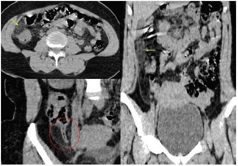
Figure 1: Shuttle sign shown in axial and coronal plan (Yellow arrow) with normal appendix (Red circle).
Unenhanced CT images show a well-circumscribed ovoid mass in the coeco-appendicular junction or adjacent to the left colon, containing fat surrounded by hyperdense ring in a “shuttle” shape, with fat stranding of the visceral peritoneum surrounding the epiploic appendage. A central high density dot can also be seen as a “dot sign”, which is highly evocative of venous thrombus within an inflamed epiploic appendage [3].
A correct identification of appendagitis is crucial as it mimics acute abdominal diseases for which surgery is required.
References
- Nhamoucha Y, Bouabdellah Y. A rare cause of acute abdomen: epiploic appendagitis. Pan Afr Med J. 2018; 30: 267.
- Charifi Y, Lamrani Y, Chbani L, Maaroufi M, Alami B. Acute abdomen in adult revealing unusual complicated epiploic appendagitis: A case report. Int J Surg Case Rep. 2020; 75: 112-116.
- Ajay K Singh, Debra A Gervais, Peter F Hahn, James Rhea, Peter R. CT Appearance of Acute Appendagitis. Mueller American Journal of Roentgenology. 2004; 183: 1303-1307.