Clinical Image
Austin J Radiol. 2023; 10(2): 1216.
Mycotic Cerebral Aneurysm: A Rare Complication of Infective Endocarditis
Kaouthar Sfar*; Rachida Chehrastane; Kaoutar Maslouhi; Fatima Zahra Laamrani; Jroundi Laila
Emergency Radiology Department of the University Hospital Ibn, Sina Rabat, Morocco
*Corresponding author: Sfar K Emergency Radiology Department of the University Hospital Ibn, Sina Rabat, Morocco. Email: sfarkaouthar5@gmail.com
Received: May 10, 2023 Accepted: June 02, 2023 Published: June 09, 2023
Clinical Image
Intracranial mycotic aneurysm or Infected Aneurysm (IA) is rare and accounts for 2-4 % of neurologic complications of infective endocarditis [1]. The terminology «mycotic aneurysm» was used for the first time by William Osler in Gulstonian Lectures of 1885, it refers to a dilatation of an arterial wall caused by any infectious etiology [2]. It is a serious clinical condition that carries a significant morbidity with up to 80% mortality rates when reptured [3]. Therefore, an early and rapid diagnosis is critical for timely treatment to optimize patient outcome.
In infective endocarditis, a hematogenous spread of septic emboli leads to the developement of arteritis that causes weakening of the vessel wall with subsquent contained rupture and formation of the pseudoaneurysm. The mycotic aneurysm can rupture resulting in intracebral or subarachnoid hemorrhage [1,4].
Clinically, the mycotic aneurysm is silent until its rupture responsible for a subarachnoid or cerebral hemorrhage. In addition to symptoms related to infective endocarditis, the patient presents a sudden and severe headache, nuchal rigidity, photophobia, vision changes (intracranial hypertension), focal deficit, and consciousness disorders [1,5].
Computed Tomography Angiography (CTA), Digital Substraction Angiography (DSA), and Magnetic Resonance Angiography (MRA) are excellent imaging modalities to establish the diagnosis and describe the infected aneurysm [5]. Contrast enhanced MRA is more efficient than 3D time of flight MRA(TOF), but it cannot replace DSA which detect the IA<3mm [4,6].
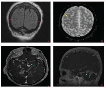
Radiographic features include focal vascular dilatation with a saclike from the vessel wall [7]. The diagnosis is established by a cluster of arguments: a systemic infection, multiple aneurysms that change in shape, size and number with arterial stenosis or occlusion close to the aneurysm [4,7,8]. MR can show other cerebral lesions: brain microbleeds, subarachnoid hemorrhage, parenchymal hemorrhage, and cerebral abscess (Figure).

Figure 1:
Treatment includes antibiotics in association with endovascular or open surgery [1].
References
- Arvin R Wali, Robert C Rennert, Jeffrey A. Steinberg, David S. Dieppa, Jeffrey S, et al. Mycotic cerebral aneurysms. Intracranial aneurysms. 2018: 663-70.
- Osler W. The Gulstonian Lectures, on Malignant Endocarditis. Br Med J. 1885; 1: 522-526.
- SV Avallone, AS Levy, RM Starke. A rare case of Streptococcus anginosus infectious intracranial aneurysm: Proper management of a poor prognosis. Surg Neurol Int. 2021; 12: 487.
- Lee WK, Mossop PJ, Little AF, Fitt GJ, Vrazas JI, et al. Infected (Mycotic) Aneurysms: Spectrum of Imaging Appearances and Management. RadioGraphics. 2008; 28: 1853-1868.
- R Sonnevillea, I Kleinb, L Bouadmaa, B Mourvilliera, B Regniera, et al. Neurologic complications of infective endocarditis. Reanimation. 2009; 8: 547-55.
- E Unlu, B Cakir, B Gocer, N Tuncbilek, M Gedikoglu. The role of contrast-enhanced MR angiography in the assessment of recently ruptured intracranial aneurysms: a comparative study. Neuroradiology. 2005; 47: 780-791.
- G Shen, X Shen, G Zhang, A Lerner, B Gao. Imaging of cerebrovascular complications of infection. Quant Imaging Med Surg. 2018; 8: 1039-1051.
- Kuo I, Long T, Nguyen N, Chaudry B, Karp M, et al. Ruptured Intracranial Mycotic Aneurysm in Infective Endocarditis: A Natural History. Case Reports in Medicine. 2010; 168408: 1-7.