
Clinical Image
Austin J Radiol. 2023; 10(3): 1217.
Cortical Laminar Necrosis: Imaging Features and Etiological Oorientation
Nourrelhouda Bahlouli, MD*; Fatima Chait, MD; Nazik Allali, PhD; Siham El Haddad, PhD; Latifa Chat, PhD
Pediatric Teaching Hospital, Department of Radiology, Mohammed V University, Rabat, Morocco
*Corresponding author: Nourrelhouda Bahlouli Pediatric Teaching Hospital, Department of Radiology, Mohammed V University, Rabat, Morocco. Email: nourrelhoudabahlouli14@gmail.com
Received: May 29, 2023 Accepted: June 23, 2023 Published: June 30, 2023
Clinical Image
Cortical laminar necrosis corresponds to neuronal ischemic remodeling with gliosis and accumulation of lipid-rich macrophages secondary to various etiologies: hypoxia, prolonged arterial hypotension, hypoglycemia, status epilepticus or arrest. Diffusion and T2 (FLAIR) show cortical and basal ganglia hypersignal from the first 24 hours. T1 shows cerebral edema during the first two weeks and a characteristic spontaneous cortical and basal ganglia hypersignal delayed and transient (3rd week). On T2*, the cortical lesions do not show any asignal, which makes it possible to differentiate them from a hemorrhagic transformation. Cortical contrast can be observed from the first weeks and for several months (up to 9 months in case of focal ischemia) and translates a rupture of the blood-brain barrier. In CT, hypodense and hyperdense gyriform density abnormalities are seen depending on the time.
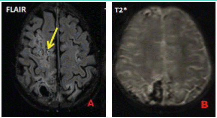
Figure A and B: MRI axial Flair and T2* slices showing cortical gyriform flair hypersignal without asignal on T2* sequences.
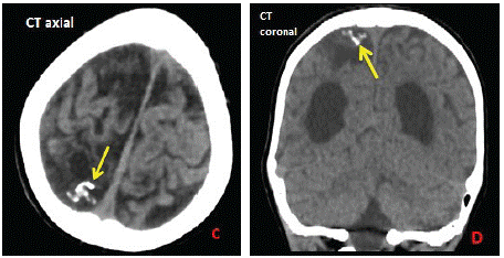
Figure C and D: CT axial section and coronal reconstruction showing the appearance of lesions described on MRI of the same patient corresponding to sequellar calcified gyriform hyperdensity.
Differential diagnoses can be made with: Sturge-Weber-Krabbe syndrome, encephalitis, PRES, arteriovenous malformation.
Cerebral cortical laminar necrosis might be another clue for predicting the poor prognosis in an individual with neuronal aggression.