Case Report
Austin J Radiol. 2023; 10(3): 1218.
High-Flow Indirect Carotid-Cavernous Fistula with Transarterial and Transvenous Endovascular Treatment
Alexandre Mello Savoldi, MD*; Mayara Thays Beckhauser, MD, MSc; Thiago Vilela Calzada Machado, MD; Gelson Luiz Koppe
Hospital Universitário Cajuru, Pontifícia Universidade Católica do Paraná – Curitiba/PR, Brazil
*Corresponding author: Alexandre Mello Savoldi Hospital Universitário Cajuru, Pontifícia Universidade Católica do Paraná – Curitiba/PR, Brazil. Email: alesavoldi94@gmail.com
Received: June 19, 2023 Accepted: July 19, 2023 Published: July 26, 2023
Abstract
Male, 39 years old, with a progressive headache for seven months, progressive right exophthalmos, right chemosis and conjunctival edema, pulsatile tinnitus and carotid bruit in the right frontotemporal region. Cerebral magnetic nuclear angioresonance showed multiple anomalous vascular structures near the sphenoparietal sinus, pterygoid plexus, superior and inferior orbital fissures, all on the right, with swelling of the basilar plexus. A diagnostic cerebral and cervical arteriography was performed, which showed a high-output indirect Carotid-Cavernous Fistula (CCF), nourished mainly by multiple dysplastic dural branches originating from the mandibular and pterygopalatine segments of the internal maxillary artery and secondarily by the inferolateral trunk. There was hypertensive venous drainage anteriorly through the ecstatic superior and inferior ophthalmic veins, corresponding angular and facial veins, and posteriorly to both cavernous sinuses, basilar plexus, superior petrosal sinuses, and internal jugular veins (Type D, Barrow Classification). The patient underwent arterial embolization with hystoacril and lipiodol in branches of the right internal maxillary artery and venous embolization with platinum micro coils with occlusion of the fistula. He evolved well with the improvement of symptoms, being discharged from the hospital and scheduled for outpatient follow-up.
The CCFs are an abnormal shunt between arteries and veins within the cavernous sinus, mainly causing a change in the distribution of cerebro-orbital blood flow. The CCFs can be spontaneous or traumatic, with high or low flow. Due to the anatomical differences, Barrow’s classification categorizes CCF according to the angiographic pattern of arterial flow, as type A or direct (direct communication between the cavernous segment of the internal carotid artery and the cavernous sinus) and type B-D or indirect (indirect communication between branches of the internal and/or external carotid arteries and the cavernous sinus). Symptoms result from venous hypertension in the venous sinuses and reversal of flow to the veins. Due to the anatomical complexity of CCF, treatment is a challenge, and the goal is to interrupt the fistulous communications and decrease the pressure in the cavernous sinus. Endovascular treatment is the first choice with occlusion of the fistulous area using materials such as a detachable balloon, coils, liquid embolic agents or covered stents. Type A fistulas are usually treated transarterial, and type B–D, by transarterial and/or transvenous embolization.
Keywords: Carotid-cavernous fistula; Cavernous sinus; Endovascular treatment
Introduction
Cavernous Carotid Fistulas (CCFs) represent abnormal arteriovenous communication between the cavernous sinus and the carotid arterial system (internal and external carotid arteries) [1-3]. They can be characterized by high or low flow, direct or indirect (dural), traumatic or spontaneous. The clinical manifestations can vary according to the type of FCC and the venous drainage pattern [1-4].
Barrow classification categorizes the CFF into direct (type A) and indirect (types B, C and D) types. Type A is a direct high-flow shunt between the internal carotid artery and the cavernous sinus. Type B is a dural shunt between meningeal branches of the internal carotid artery and the cavernous sinus. Type C is a dural shunt between meningeal branches of the external carotid artery and the cavernous sinus. Type D is a dural shunt between meningeal branches of the internal and external carotid arteries and the cavernous sinus [5].
Direct FCCs have a rapid flow. They are more common in young men, frequently caused by traumatic insult, by rupture of an internal carotid artery aneurysm within the cavernous sinus, associated with an underlying connective tissue disorder (Ehlers-Danlos syndrome type IV), or iatrogenic intervention [1-4].
Indirect FCCs usually have a low flow and are more common in middle-aged women, with 25% of these fistulas being spontaneous; other causes include hypertension, fibromuscular dysplasia, Ehlers-Danlos type IV, and dissection of the internal carotid artery [1-3].

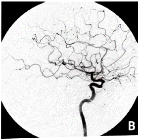
Figure 1: Male, 39 years old, with a progressive headache for seven months, right exophthalmos, right chemosis and conjunctival edema, pulsatile tinnitus and carotid bruit in the right frontotemporal region. Diagnostic cerebral and cervical arteriography: right internal carotid artery injection, arterial phase by anteroposterior view (A) and lateral view (B); right external carotid artery injection, arterial phase by anteroposterior view (C) and venous phase by anteroposterior view (D) which showed a high-output indirect carotid-cavernous fistula (CCF). The feed is mainly by multiple dysplastic dural branches originating from the mandibular and pterygopalatine segments of the internal maxillary artery and secondarily by the inferolateral trunk, with hypertensive venous drainage anteriorly through the ecstatic superior and inferior ophthalmic veins, corresponding angular and facial veins, and posteriorly to both cavernous sinuses, basilar plexus, superior petrosal sinuses, and internal jugular veins (E-H).

Figure 2: The patient underwent arterial embolization with hystoacril and lipiodol in branches of the right internal maxillary artery and venous embolization with platinum micro coils with occlusion of the fistula. Final arteriography: right internal carotid artery injection, arterial phase by anteroposterior view (A) and lateral view (B); right external carotid artery injection, arterial phase by lateral view (C); venous phase by anteroposterior view (D) and lateral view (E); and final angiogram demonstrates coil embolization of the CCF (F).
The FCCs symptoms are related to size, duration, location, presence of venous or arterial collaterals, and venous drainage. They include headache, intracranial haemorrhages (subarachnoid and subdural), cephalic bruit, trigeminal nerve dysfunction and epistaxis, resulting from venous hypertension in the dura mater sinuses. The most frequent ocular signs and symptoms are exophthalmos, proptosis, conjunctival chemosis (red eye), ophthalmoplegia, ocular pain, increased intraocular pressure and decreased visual acuity, resulting from the reversal of blood flow to the ophthalmic veins due to overload and dilation of the cavernous sinus [3-6].
Treatment depends on flow velocity through the fistula, arterial supply, and venous drainage routes. Endovascular intervention or stereotactic radiosurgery can be performed when invasive treatment is necessary. Modern endovascular techniques offer the ability to treat CCF with low morbidity successfully [7,8].
In this case report, we aimed to describe a case report of high-flow complex indirect FCC that underwent endovascular treatment via transarterial and transvenous.
Case Report
Male, 39 years old, with a progressive headache for seven months, progressive right exophthalmos, right chemosis and conjunctival edema, pulsatile tinnitus and carotid bruit in the right frontotemporal region. Cerebral magnetic nuclear angioresonance showed multiple anomalous vascular structures near the sphenoparietal sinus, pterygoid plexus, superior and inferior orbital fissures, all on the right, with swelling of the basilar plexus. A diagnostic cerebral and cervical arteriography was performed, which showed a high-output indirect Carotid-Cavernous Fistula (CCF), nourished mainly by multiple dysplastic dural branches originating from the mandibular and pterygopalatine segments of the internal maxillary artery and secondarily by the inferolateral trunk. There was hypertensive venous drainage anteriorly through the ecstatic superior and inferior ophthalmic veins, corresponding angular and facial veins, and posteriorly to both cavernous sinuses, basilar plexus, superior petrosal sinuses, and internal jugular veins (Type D, Barrow Classification).
The patient underwent arterial embolization with cyanoacrylate (hystoacril) and lipiodol in branches of the right internal maxillary artery and venous embolization with platinum micro coils with occlusion of the fistula.
He evolved well with improving symptoms, being discharged from the hospital and scheduled for outpatient follow-up.
Discussions
We present a case in which our patient presented an indirect high-flow FCC, with proptosis, conjunctival chemosis and cranial bruit at the frontotemporal region. The sudden and intense headache and periorbital edema corroborate the diagnostic hypothesis. Since the patient did not suffer any trauma, added to the absence of paralysis of the extrinsic eye muscles, amaurosis or subarachnoid hemorrhage, this leads us to believe that it fits into the category of spontaneous CCF.
Cerebral arteriography remains the gold standard for the FCC classification, diagnosis and therapy. Angiographic evaluation defines the size and location of the CCF, differentiation of direct from indirect lesions, high or low flow, presence of an associated aneurysm, assessment of global cortical arterial circulation and collateral flow through the circle of Willis and identification of venous drainage characteristics. Angiography performed on our patient showed the presence of Barrow Type D high-flow indirect right FCC, with multiple arterial inputs and drainage veins. These indirect CCFs are treated if symptoms are intolerable or vision is threatened [3-8]. Our patient's decision to treatment was due to the fistula presenting high flow with progressive symptoms.
Endovascular treatment is the first choice, with occlusion of the FCC fistulous area using materials: detachable balloons, coils, liquid embolic agents, or a combination of these. Type A FCC is usually treated by transarterial approach, with the objective of occluding the direct communication between the internal carotid artery and the cavernous sinus, preserving the permeability of the internal carotid artery and its branches. Type B–D FCC often resolve spontaneously, low-risk cases may be managed conservatively, but when necessitating treatable, the approach is by transarterial and/or transvenous embolization. The aim of treatment is to stop the fistulous communications and decrease the pressure in the cavernous sinus, occluding the arterial branches that supply the fistula (transarterial embolization) or, more commonly, occluding the cavernous sinus that houses the fistulous communications (transvenous embolization). When transvenous approaches are not feasible due to vessel tortuosity, sinuous venous thrombosis or occlusion, a direct orbital approach to the cavernous sinus with fluoroscopic guidance may be considered [6-8].
In this reported case, transarterial embolization with embolic material was performed, obliterating an anatomically complicated FCC. Despite the increasing success of transarterial procedures, a transvenous approach via the inferior petrosal sinus, superior petrosal sinus, basilar plexus, pterygoid plexus, superior orbital vein, or inferior ophthalmic vein is still preferred for most dural CCF requiring treatment [6-8]. Due to the need for occlusion of the fistulous area, the patient also underwent venous embolization with platinum microcoils. In this regard, the inferior petrosal sinus is the first-line approach, as it is the simplest and shortest route to the cavernous sinus. Advances in endovascular technology, including the development of microcatheters of varying stiffness and guidewires, have increased the feasibility of this approach.
Conclusion
Our patient had indirect high-flow CCF with multiple arterial inputs and drainage veins, which marked attention due to the complexity and difficulty of accessing the fistulous area. CCFs are rare but treatable causes of orbital injury and vision loss. The FCC etiology must be investigated, reaffirming the importance of a detailed anamnesis since the symptoms are usually insidious, especially in dural fistulas. Early intervention in reflux into cortical veins and dilation of the ophthalmic vein is essential to avoid complications such as intracranial hemorrhage and progressive visual loss. Although a watchful approach is reasonable in many patients with a dural FCC, treatment is sometimes necessary to avoid long-term sequelae. Successful closure can usually be achieved with acceptable morbidity using current endovascular techniques for patients with high-flow fistulas and those with cortical venous drainage.
References
- Henderson AD, Miller NR. Carotid-cavernous fistula: current concepts in aetiology, investigation, and management. Eye (Lond). 2018; 32: 164-72.
- Reynolds MR, Lanzino G, Zipfel GJ. Intracranial dural arteriovenous fistulae. Stroke. 2017; 48: 1424-31.
- Alexander MD, Darflinger R, Cooke DL, Halbach VV. Cerebral arteriovenous fistulae. Handb Clin Neurol. 2021; 176: 179-98.
- Lasjaunias P, Chiu M, ter Brugge K, Tolia A, Hurth M, Bernstein M. Neurological manifestations of intracranial dural arteriovenous malformations. J Neurosurg. 1986; 64: 724-30.
- Barrow DL, Spector RH, Braun IF, Landman JA, Tindall SC, et al. Classification and treatment of spontaneous carotid cavernous sinus fistulas. J Neurosurg. 1985; 62: 248-56.
- Oushy S, Borg N, Lanzino G. Contemporary management of cranial dural arteriovenous fistulas. World Neurosurg. 2022; 159: 288-97.
- Zanaty M, Chalouhi N, Tjoumakaris SI, Hasan D, Rosenwasser RH, et al. Endovascular treatment of carotid-cavernous fistulas. Neurosurg Clin N Am. 2014; 25: 551-63.
- Gemmete JJ, Ansari SA, Gandhi DM. Endovascular techniques for treatment of carotid-cavernous fistula. J Neuroophthalmol. 2009; 29: 62-71.