Clinical Image
Austin J Radiol. 2023; 10(4): 1223.
Solitary Bone Plasmacytoma of Spine
Benmoussa Meryem*; Chait Fatima; Zahi Hiba; Imrani Kawtar; Moatassim Billah Nabil; Nassar Itimade
Department of Central Radiology, Ibn Sina Hospital, Mohammed V University, Morocco
*Corresponding author: Benmoussa Meryem Department of Central Radiology, Ibn Sina Hospital, Mohammed V University, Morocco. Tel: +212659209483 Email : benmoussa.mer93@gmail.com
Received: November 09, 2023 Accepted: December 21, 2023 Published: December 28, 2023
Clinical Image
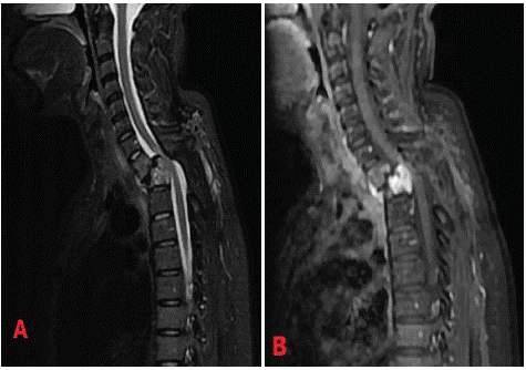
A 54-year-old male presented a chronic intense back pain that progressed to paraplegia and urinary disorders. His lateral spinal x-ray showed osteolytic destruction of T1 and T2 vertebrae with intervertebral space stenosis. MRI detected a single malignant osteolytic process of the spine involving T1 and T2 with a pathologic fracture leading to segmental kyphosis. Epidural soft tissue mass with typical curtain sign was causing spinal cord compression Figure 1.

Figure 1: A and B : Sagittal magnetic resonance imaging reveals an epidural mass involving T1 and T2 vertebral body causing spinal cord compression and pathologic fracture leading to segmental kyphosis.
The patient underwent emergency surgery, undergoing posterior laminectomy and stabilization of the dorsal spine combined with bone biopsy. Histology revealed a vertebral plasmacytoma.
Solitary bone plasmacytoma is a rare tumor, representing 7% of myelomas. It’s considered to be a solitary form of multiple myeloma [1]. It’s an malignant disease, which is defined by the presence of a single osteolytic lesion due to monoclonal plasma cell infiltration [2]. Most of patients are treated with radiotherapy, while others require surgery.
Disclosure of Interest
The authors declare that they have no conflicts of interest concerning this article.
References