Rapid Communication
Austin J Radiol. 2024; 11(1): 1224.
Coronary Artery and Aortic Valve Calcification Quantification in CT Chest Studies of Lung Cancer Screening
Girgis M*; Gutierrez M; McCreavy D; Radike M; Fairbairn T
Liverpool Heart & Chest Hospital, Liverpool, United Kingdom
*Corresponding author: Mina Girgis Liverpool Heart & Chest Hospital, Liverpool, United Kingdom. Email: minamaher195@gmail.com
Received: December 26, 2023 Accepted: January 20, 2024 Published: January 27, 2024
Abstract
Reporting Coronary Artery Calcification (CAC) and Aortic Valve Calcification (AVC) on non-gated, non-contrast CT scans is recommended in the guidelines. This study aimed to assess the adherence guidelines in reporting incidental CAC and AVC present in the scans. It also aimed to assess the agreement between visual assessment and Agatston Score (AS) evaluating the severity of CAC and AVC in lung cancer screening chest CTs scans. This retrospective study included 100 patients’ low dose, non-gated, non-contrast CT scans. The scans were retrieved from the targeted lung health check program. The visual assessment of CAC and AVC was done by multiple reporters participating in the screening program. It was interpreted as none, mild, moderate or severe. The Agatston score, a reliable precursor of CAC and AVC was calculated using Siemens SyngoVia and interpreted as none, mild (<100), moderate (101-300) or severe (>300). The two scoring systems were compared to each other. CAC was present in 81 % of the cases and there was a comment on it in every report. There was a moderate agreement between the visual assessment of CAC and AS. Weighted kappa score was 0.516. AVC was reported in 4 cases of the 100 cases. It was present in only one case of them after reviewing the Agatston score of all the cases. AVC was present in 6 cases. It was reported in one case and missed in five cases. The reported case was with moderate AVC, and the missed cases were all with mild AVC.
Keywords: Chest CT; Cardiovascular disease; Coronary artery calcium
Introduction
Atherosclerotic cardiovascular disease is the leading cause of death in the UK. Coronary heart disease causes almost 66000 deaths each year, roughly 180 people each day, or one death every eight minutes [1]. Approximately one in eight men and one in fourteen women die from coronary heart disease, which makes primary prevention crucial to prevent events leading to death or long-term disability. In the UK, primary prevention with lifestyle modification is indicated in all patients with any cardiovascular risk factor [2]. When the 10-year risk of future cardiovascular events is 10 % or more, preventive therapy with statins is indicated. The individual cardiovascular risk is assessed using models that consider demographics, clinical and biochemical factors [3-5]. However, coronary calcium score has proven in multiple studies to outperform the prediction power of traditional risk prediction models [6]. In a recent multicentric study including more than 60.000 patients higher coronary calcium increased cardiovascular, and all-cause mortality regardless of risk factors burden [7].
The gold standard for assessment of Coronary Artery Calcification (CAC) is the Agatston score, which is based on an ECG-gated, non-contrast enhanced CT covering the heart. 3mm thick slices without overlap are acquired in end-diastole. Coronary calcification is defined as areas with >130 Hounsfield Units (HU) and at least 1mm2 (=3 consecutive pixels) [8]. The score is then calculated by adding the weighted sum of lesions (higher density lesions score higher) and classified in categories linked to the risk of future cardiovascular events. Similar, but slightly different classifications exist. In this paper we followed the one endorsed by the Society of Cardiovascular Computer Tomography and the Society of Thoracic Radiology in 2016: Agatston score 0 = no CAC (very low risk), 1-99, mild CAC (mildly increased risk), 100-299= moderate CAC (moderately increased risk), =300= moderate to severe CAD (moderately to severely increased risk) [9].
Despite the absence of ECG gating, chest CTs are a perfect chance to provide opportunistic risk stratification to patients being scanned for multiple reasons. With this in mind, a joint statement by the BSCI/ BSCCT and BSTI has recommended reporting the degree of coronary calcium in all thoracic scans. They recommend the heart to be reviewed on all CT scans where it is covered, and the presence of coronary artery and aortic valve calcification identified and quantified using simple visual quantifications (none, mild, moderate severe).
So far, various studies have evaluated the agreement between visual analysis and CAC scoring on non-ECG gated CTs in comparison with CAC scoring using ECG-gated CTs. Their results show good agreement in the identification of CAC between ECG-gated Agatson score, non-ECG gated Agatson score and visual assessment [10]. All these studies used a limited number of reviewers and/or a semiquantitative scale when visual evaluation was used.
In this descriptive study, we aim to assess agreement between calcium score and visual assessment on low dose lung cancer screening chest CTs. Studies have been reported by multiple readers, as part of their daily clinical practice, without guidance on how to quantify coronary calcification. All reports within the UK Targeted Lung Health Check program are structured, which guarantees adherence to the recommendation of reporting coronary artery calcification. Additionally, we have assessed the adherence to the recommendation of identifying and quantifying aortic valve calcification.
Methods
Institutional Review Board approval was waived by our Research and Innovation Department, which classified the study as Service Evaluation. A retrospective, single centre study was performed. One hundred low dose chest CTs from the lung cancer screening program acquired between March 2022 and June 2023 were filtered on PACS and reviewed. The only inclusion criteria were belonging to the Targeted Lung Health project. Patients from across the UK aged from 55-74 who have a positive smoking history receive an invitation by post to have a low dose CT scan. Patients without significant findings are receive a follow up invitation in 2years. Shorter term follow ups or referral for further investigations can be done when appropriate. Two exclusion criteria were applied: patients with previous coronary intervention and follow up scans.
All studies were acquired following NHS England recommended protocol for lung cancer screening [11]. All patients lay supine with the arms above their head and thorax centred on the table. Images were obtained in maximal inspiration following a low dose volumetric protocol. Automatic dose modulation was implemented to adapt the kilovoltage (Kv) and milliamperage (mAs) to body habitus with the objective of keeping total dose below 2mSv. Thin axial slices were reconstructed using lung and soft tissue windows. Additionally, a sagittal reconstruction was obtained for assessment of the spine (e.g., incidental compression fractures).
Images were interpreted and reported by multiple thoracic radiologists participating in the reginal lung cancer screening program, without particular guidance on how to report coronary or aortic valve calcification. A single operator retrospectively assessed the radiological reports and quantified coronary artery calcification (Agatston score) using Siemens SyngoVia. Visual analysis identified four degrees of coronary calcification: none, mild, moderate and severe. In consequence, the Agatston score levels were reclassified to 0=none, 1-99= mild, 100-299=moderate and =300=severe.
Apart from the coronary artery evaluation, it was also assessed if the there was a comment on aortic valve calcification. As with coronary calcification the visual scoring system was none, mild, moderate or severe. The aortic valve calcium score was calculated on SyngoVia.

The collected data was stored on Microsoft Excel and the statistical analysis performed using R. Agreement was measured by percentage agreement (overall and between categories) and weighted Kappa. Linear and quadratic weighting were used for completion.
Results
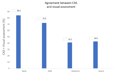
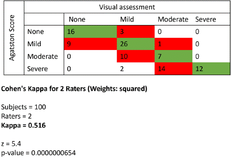
The study included 60 women and 40 men with a mean age of 66.4 years. Coronary calcification was present in 81 cases. It was reported in all the cases. Even if CAC was absent, it was mentioned in the report. In 16/19 (84%) of patients without coronary calcification and 26/36 (72.2%) with mild CAC the calcium score and visual evaluation agreed. In 7/17 (41.1%) of patients with moderate CAC and 12/28 (42.8%) of patients with severe CAC the two scores agreed.

Weighted Kappa test showed moderate agreement between the two scoring systems (k=0.516). There were 39 cases that showed discrepancies between the visual scoring and Agatston scoring. Out of the 39 cases with discrepancies between visual scoring and Agatston, 9 cases with mild scoring were reported as having no CAC visually. 3 cases without CAC were reported as having mild CAC visually. 10 cases with moderate scoring were reported as having mild CAC visually. 14 cases with severe CAC were reported as having moderate CAC visually and 2 cases with severe CAC were reported as having mild CAC visually.

Regarding the results of the aortic valve, out of the 100 reports, 4 reports had a comment on presence or absence of aortic valve abnormalities (3 AVC, 1 aortic valve prosthesis). Out of the 3 reported with AVC, 1 had AVC upon review of Agatston scoring (1 true positive and 2 false positive). A total of 6/100 had AVC (5 mild and 1 moderate), with 5 of them with mild AVC not reported and one case with moderate AVC reported.
Discussion
The screening of CAC in national lung cancer screening programs has shown improving numbers in terms of reducing mortality [12]. This is supported by the fact that lung cancer and CAC share several risk factors, one of them is the past or current smoking history. Both also tend to occur in the same age group (55-74 years). The concomitant screening for CAC on the same low dose CT scan performed in the lung screening program spares the patient extra radiation and demonstrated cost effectiveness in comparison to other screening tests for CAC [13].
Our study aimed to evaluate the adherence to the latest recommendations of the BSCI regarding reporting incidental CAC and AVC in routine CT chest scans. The visual evaluation of CAC was done in 100% of the scans and the practice showed high adherence to the previously mentioned recommendations [9]. In a similar retrospective study of visual ordinal scoring of coronary artery calcium on contrast-enhanced and non-contrast chest CTs, Camila et al. found that CAC was mentioned in only 37% (53/144) of clinical reports of non-contrast chest CTs [14]. The higher percentage of visual evaluation of CAC in our study is attributed to the structured reporting software the radiologist participating in the targeted lung health check scans use. The software was designed to draw the attention of the reporter to report and visually estimate CAC.
However, in our study this wasn’t the case for reporting AVC. This was probably because it wasn’t widespread in the screening sample and was present in only 6 % of the cases. Even though AVC wasn’t prevalent, there was a trend of discrepancy in reporting it.
Our study aimed to evaluate the diagnostic performance of reporting CAC and AVC on low dose non-gated non-enhanced CT chest scans. It also aimed to evaluate the agreement of visual assessment and Agatston scoring of CAC and AVC. Agreement between visual and Agatston scoring was higher in patients with no or mild CAC. Whereas this agreement decreased substantially in moderate and severe CAC which conditions an overall moderate agreement in all cohort.
In a similar retrospective study of 260 patients’ CT scans, Camila et al. found an excellent interobserver agreement in the visual ordinal CAC assessment on non-contrast, non-gated CT scans with a kappa value of 0.95 [14]. In this study the visual assessment of CAC was done by a single cardiac radiologist who assigned each CT chest scan a single overall score reflecting the presence and severity of CAC assessed visually in a global fashion across all epicardial coronary arteries using a previously validated ordinal scale: none (no CAC visualized), mild (isolated speckles of CAC in any coronary artery), moderate (more than mild, less than severe), or severe (at least one coronary segment with contiguous CAC or with extensive or diffuse CAC). To evaluate the interobserver agreement in this study, a second cardiac radiologist who was blinded to the results of the first observer evaluated a random subset of 50 chest CT examinations. In our study the visual evaluation of CAC was conducted by multiple reporters (not a single reporter) participating in the targeted lung health check program. Most of the reporters who participated are chest radiologists and the visual assessment was done without using any semi-quantitative methods and without guidance on how to quantify coronary artery calcification.
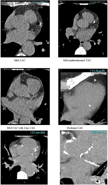
A coronary artery calcium score of zero is the most important negative risk predictor in asymptomatic and symptomatic patients [15]. Therefore, the greatest concern of low dose, non-gated, non-contrast CT scans is the underestimation of coronary calcium due to inability to detect small calcifications [16]. In our study, 9 % of the patients were misclassified as zero Agatston score despite having calcium on their scans. Underestimating CAC was not only present in cases with mild CAC, but it was also present in cases with moderate and severe CAC.
The findings of our study have an important implication which is routine reporting of CAC in low dose non-contrast non-enhanced CT chest examinations could identify many patients with previously unknown CAC who might benefit from preventive treatment.
Conclusions
In this study, there was a moderate agreement between visual and the Agatston scoring systems. Agreement between visual and Agatston scoring was higher in patients with no or mild CAC whereas this agreement decreased substantially in patients with moderate and severe CAC which conditions an overall moderate agreement in all cohort. While AVC was not prevalent, its reporting was low with a trend for discrepancy. Standardization of visual scoring and inclusion of aortic valve reporting are recommended in the lung cancer screening chest CT proformas.
Author Statements
Conflicts of Interest
The authors have no conflicts of interest to declare.
References
- UK factsheet. British Heart Foundation. 2023.
- Stewart J, Addy K, Campbell S, Wilkinson P. Primary prevention of cardiovascular disease: updated review of contemporary guidance and literature. JRSM Cardiovasc Dis. 2020; 9: 2048004020949326.
- Risk estimation and the prevention of cardiovascular disease. Sign. 2017; 149.
- ’Cardiovascular disease: risk assessment and reduction, including lipid modification,’ NICE guideline; 2014.
- Lim HY, Burrell LM, Brook R, Nandurkar HH, Donnan G, Ho P. The need for individualized risk assessment in cardiovascular disease. J Pers Med. 2022; 12: 1140.
- Budoff MJ. Expert review on coronary calcium. Dovepress. 2008.
- Grahndhi GR. Interplay of coronary artery calcium and risk factors for predicting CVD/CHD mortality: the CAC consortium Journal of the American College of Cardiology. 2019.
- Hecht HS. 2016 SCCT/STR guidelines for coronary artery calcium scoring of noncontrast noncardiac chest CT scans: A report of the Society of Cardiovascular Computed Tomography and Society of Thoracic Radiology. J Cardiovasc Comput Tomogr. 2016.
- Williams MC. Reporting incidental coronary, aortic valve and cardiac calcification on non-gated thoracic computed tomography, a consensus statement from the BSCI/BSCCT and BSTI. Br J Rad. 2020.
- Pelandré GL, Sanches NMP, Nacif MS, Marchiori E. Detection of coronary artery calcification with nontriggered computed tomography of the chest. Radiol Bras. 2018; 51: 8-12.
- Standard protocol prepared for the Targeted Lung Health Checks Programme. NHS England. 2022.
- Fan L, Fan K. Lung cancer screening CT-based coronary artery calcification in predicting cardiovascular events: a systematic review and meta-analysis. Medicine. 2018; 97: e10461.
- Waltz J, Kocher M, Kahn J, Dirr M, Burt JR. The future of concurrent automated coronary artery calcium scoring on screening low-dose computed tomography. Cureus. 2020; 12: e8574.
- Fresno CU. Visual ordinal scoring of coronary artery calcium on contrast-enhanced and noncontrast chest CT: A retrospective study of diagnostic performance and prognostic utility. Am J Roengenology. 2022; 219: 569-578.
- Blaha MJ, Cainzos-Achirica M, Greenland P, McEvoy JW, Blankstein R, Budoff MJ, et al. Role of coronary artery calcium score of zero and other negative risk markers for cardiovascular disease: The Multi-Ethnic Study of Atherosclerosis (MESA). Circulation. 2016; 133: 849-58.
- Dobrolinska MM. Performance of visual, manual, and automatic coronary calcium scoring of cardiac 13N-ammonia PET/low dose CT. J Nucl Cardiol. 2022.