Clinical Image
Austin J Radiol. 2024; 11(2): 1232.
Imaging Features of Bilateral Involvement in Sturge-Weber Syndrome
Rachida Chehrastane*; Kaouthar Sfar; Sara Essetti; Sanae Jellal; Siham El Haddad; Nazik Allali; Latifa Chat
Department of Pediatric Radiology, Ibn Sina University Hospital Center, Rabat, Morocco
*Corresponding author: Rachida Chehrastane Department of Pediatric Radiology, Ibn Sina University Hospital Center, Rabat, Morocco. Email: chehrastane.rachida@gmail.com
Received: April 10, 2024 Accepted: May 03, 2024 Published: May 10, 2024
Clinical Image
Sturge-Weber syndrome is a neurocutaneous phacomatosis due to an embryonic malformation of the foetal vascular system. It is characterised by two types of malformations: a congenital facial wine stain and a leptomeningeal capillary-venous angioma, associated with ocular manifestation [1-3] Generally located homolaterally in the classic form, although bilateral involvement is possible but remains rare [4].
The diagnosis of SWS relies massively on neuroimaging, specifically Magnetic Resonance Imaging (MRI), which is critical for identification abnormalities that precede neuro-ocular complications.
The diagnosis is usually obvious because of the cutaneous angioma, involving the ophthalmic branch (V1) of the trigeminal nerve. the main neurological manifestations are dominated by epileptic seizures, affecting between 75% and 90% of patients. Around half of patients also present with a motor deficit. associated with convulsions, headaches or migraines. Although psychiatric disorders and mental retardation have been reported, they remain rare [1,2]. Radiological findings can be seen on both CT scan and brain MRI, but the MRI is better than the CT scan, at detecting early signs, sometimes even before clinical symptoms appear [5,6] including:

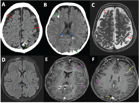
    Figure 1: CT and MRI images in a 10-year-old patient with bilateral sturge-weber syndrome.

Non injected CT sections (A, B) show bilateral frontoparietal atrophy (red arrows), right frontal and left parietal calcifications (green arrows), and choroid hypertrophy (blue arrows)

Axial T2 sequence (C) shows bilateral frontoparietal atrophy. Axial flair sequence (D) shows choroid plexus hypertrophy. Axial T1 sequence after gadolinium injection (E, F) with leptomeningeal contrast (yellow arrows) and bilateral dilatation of some intraparenchymal veins.
Focal or hemispheric cerebral atrophy
Cortical or subcortical calcifications
Hypertrophy and calcification of the choroid plexus
Choroid plexus angioma
Leptomeningeal enhancement due to congested internal veins Dilatation of trans-parenchymal veins.
References
- Habibi H, et al. Sturge-weber-krabbe syndrome: Complete form (about a case). IOSR Journal of Dental and Medical.
- Siham Alaoui Rachidi, Anas Lahlou Mimi, Amal Akammar, Youssef Lamrani Alaoui, Meriem Boubbou, Mustapha Maaroufi, Badr Alami. Sturge weber krabbe syndrome: Exceptional entity (about a case). Pan African Medical Journal. 2018.
- Sudarsanam A, Ardern-Holmes SL. Sturgee weber syndrome: From the past to the present. European Journal of Pediatric Neurology. 2013.
- Moifo B, et al. Clinical and CT scan features of bilateral sturge-weber syndrome, report of two cases. Rev. méd. Madag. 2015; 5: 586-589.
- Sherwani OA, Patra PC, Ahmad SA, Hasan S. Sturge-weber syndrome: A report of a rare case. Cureus. 2023; 15: e51110.
- Zallmann M, Mackay MT, Leventer RJ, Ditchfield M, Bekhor PS, Su JC. Retrospective review of screening for sturge-weber syndrome with brain magnetic resonance imaging and electroencephalography in infants with high-risk port-wine stains. Pediatr Dermatol. 2018; 35: 575-581.