Case Report
Austin J Radiol. 2024; 11(2): 1233.
Scar Endometriosis After Cesarean Section, A Rare Entity: Case Report
Meryem Benmoussa*; Amine Naggar; Houssein Oukili; Nazik Allali; Siham EL Haddad; Latifa Chat
Department of Radiology, Children Hospital of Rabat Mohammed V University, Morocco
*Corresponding author: Meryem Benmoussa Department of Radiology, Children Hospital of Rabat Mohammed V University, No 39, Avenue Attanoub, Hay riad, Rabat, Morocco. Tel: +212659209483 Email: benmoussa.mer93@gmail.com
Received: April 12, 2024 Accepted: May 07, 2024 Published: May 14, 2024
Introduction
Endometriosis is defined as functional endometrial glands with stroma outside the uterus, which is estimated to affect almost 10% of the reproductive age groups (15–49 years). Its incidence is estimated around 0.03–0.4%. It usually occurs around the uterus and uterine ligaments; however, extra pelvic sites, though rare, occur in previous surgical scars or cesarean scar or episiotomy scar, the lungs, brain, urinary tracts, abdominal wall, spleen, gastrointestinal tracts, and such as a previous ectopic pregnancies, hysterectomy, salpingostomy. [1] We report a rare case of parietal endometriosis occurring after Pfannenstiel scar for cesarean section, managed at Maternity Hospital of Rabat.
Case Report
A 32-year-old woman, who have two children, was seen in consultation for a painful abdominal scar and reported a subcutaneous abdominal mass, with menstrually related enlargement and pain. Her surgical history included a caesarian section two years previously, with no complications. The mass appeared six months later.
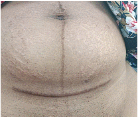
Physical examination revealed a well-healed caesarian scar (Figure 1), a found a palpable nonmobile subcutaneous mass, located under the incision scar, wich mesure approximately 5 cm.

Figure 1: Cesarean section scar with subcutaneous mass.
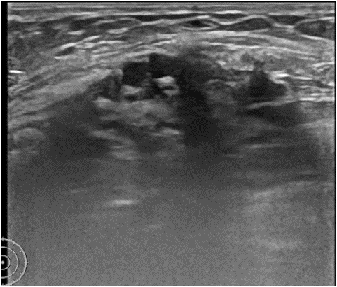
Ultrasound of the abdominal wall revealed a heterogeneous hypoechoic mass with echogenic spots inside (Figure 2).

Figure 2: Ultrasound image showing heterogenous hypoechoic mass.
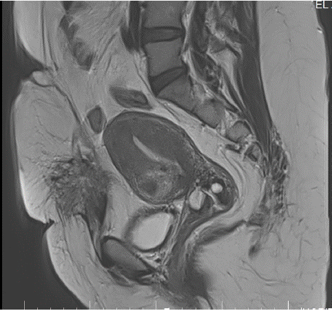
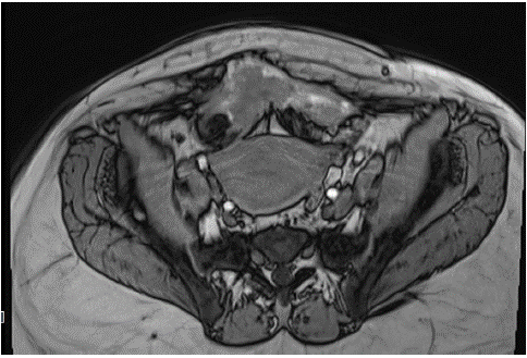
MRI confirm the presence of a parietal mass, hypointense on T1 and T2, measuring 7x5 cm, extended to the abdominal muscle and the aponevrosis (Figure 3 & 4).

Figure 3: MRI axial T1 image showing the mass in contact with the rectus abdominis muscle.

Figure 4: MRI sagittal T2 image showing the mass in contact with the rectus abdominis muscle.
The clinical history, physical examination, and imaging features led to the diagnosis of cesarean scar endometriosis.
Surgical wide excision of the mass was performed.
Anatomopathological examination confirmed the diagnosis of parietal endometriosis. Postoperatively, the patient was stable and discharged on the fourth postoperative day.
Discussion
Abdominal wall endometriosis is very related to previous history of surgery [2]. Endometriosis implants developing in the subcutaneous tissue of surgical scars occur most frequently after obstetrical and gynecological procedures, including cesarean sections, tubal ligations, cystectomies, hysterectomies and amniocenteses [3]. Cesarean Scar Endometriosis (CSE) is a rare iatrogenic disease caused by the implantation of endometrium stem cells in the incision during cesarean delivery (Pfannenstiel incision). The incidence has been estimated to be only 0.03% to 0.15% of all cases of endometriosis [1].
Many theories about the pathogenesis of CSE have been proposed, such as the implantation theory and the lymphatic or hematogenic dissemination theory [4].
The clinical presentation is variable. Pain, either cyclical or noncyclical, remained the major symptom. It’s usually intermittent and associated with the menstrual cycle, but it may be constant. The overlying skin of the scar may be hypertrophic or hyperpigmented due to the deposition of hemosiderin. However, some patients may be asymptomatic.
Ultrasound features are not specific. A subcutaneous heterogeneous mass having relatively irregular borders, predominantly hypoechoic echotexture. Internal hyperechoic echoes and vascularity may be present.
CT shows a well-defined soft tissue mass, with heterogenous post-contrast enhancement. The surrounding tissue may have a streaky appearance.
MRI is the most important and sensitive imaging modality. It confirms the diagnosis and show the exact localization. It also have an interest to search other localizations of endometriosis. Typically the lesions that can be detected with MRI are those that contain blood products, with hemorrhagic “powder burn”: lesions appear bright on T1 fat-saturated sequences, or small solid deep lesions.
The diagnosis is only confirmed by histology.
Treatment is based on large surgical excision of the lesion with clear resection margins, associated or not with hormone therapy (GnRH analogs) [5].
Conclusion
Parietal endometriosis is a rare clinical entity. It occurs most often after gynecological and obstetrical surgery. its symptomatology is variable. Clinical examination, imaging and cytology are usually efficient for the diagnosis. Treatment is based on surgical excision of the lesion with or without hormone therapy.
It is important to think about this rare diagnosis in the context of cesarian section.
Author Statements
Ethical Approval and Informed Consent
This paper did not involve any research and no ethical clearance was required. A written informed consent was obtained from the patient for the publication of this paper.
Declaration of Conflicting Interests
The author(s) declared no potential conflicts of interest with respect to the research, authorship, and/or publication of this article.
Funding
The author(s) received no financial support for the research, authorship, and/or publication of this article.
Authors Contribution
All the authors confirm responsibility for the following: study conception and design, analysis and interpretation of results, and manuscript preparation.
References
- Nominato NS, Prates LFVS, Lauar I, Morais J, Maia L, Geber S. Caesarean section greatly increases risk of scar endometriosis. Eur J Obs Gynecol Reprod Biol. 2010; 152: 83–85.
- Mistrangelo M, Gilbo N, Cassoni P, Micalef S, Faletti R, Migilietta C, et al. Surgical scar endometriosis. Surgery Today. 2014; 44: 767–772.
- Pasalega M, Mirea C, Vîlcea ID, et al. Parietal abdominal endometriosis following cesarean section. Romanian Journal of Morphology and Embryology. 2011; 52: 503–508.
- Horton JD, Dezee KJ, Ahnfeldt EP, Wagner M. Abdominal wall endometriosis: a surgeon’s perspective and review of 445 cases. Am J Surg. 2008; 196: 207–12.
- Gidwaney R, Badler R, Yam B, Hines JJ, Alexeeva V, Donovan V, et al. Endometriosis of Abdominal and Pelvic Wall Scars: Multimodality Imaging Findings, Pathologic Correlation, and Radiologic Mimics. Radiographics. 2012; 32: 2031-43.