Clinical Image
Austin J Radiol. 2024; 11(3): 1236.
Imaging Features of Choroid Plexus Hyperplasia
Doaa Alzaher*
Department of Diagnostic Radiology, Dammam Medical Complex, Dammam, Saudi Arabia
*Corresponding author: Doaa Alzaher Department of Diagnostic Radiology, Dammam Medical Complex, Dammam, Saudi Arabia. Email: dr.doaalh@gmail.com
Received: July 16, 2024 Accepted: August 02, 2024 Published: August 09, 2024
Case Presentation
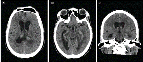
35-year-old female presented to the emergency department with 2 weeks history of headache and abnormal gait. Neurological examination revealed no sensory or motor deficits. Computer Tomography (CT) of the brain was requested in the emergency setting and showed diffuse dilatation of the lateral ventricles, third ventricle and fourth ventricle suggestive of communicating hydrocephalus. There is also diffuse enlargement of choroid plexuses in the lateral ventricles with extension into the third ventricle (Figure 1).

Figure 1: Non-enhanced CT scan of the brain. Axial (a-b) and coronal (c) planes demonstrate diffuse dilatation of the lateral ventricles, third ventricle and fourth ventricle. Diffuse enlargement of choroid plexuses in the lateral ventricles with extension into the third ventricle.
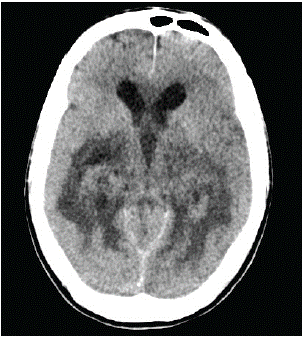
The patient was admitted and External Ventricular Drain (EVD) was inserted. Follow up scan after one day was done and showed progression of ventricular dilatation with periventricular hypodensity suggestive of Cerebrospinal Fluid (CSF) permeation in the setting of active hydrocephalus (Figure 2).

Figure 2: Axial plane of non-enhanced CT scan of the brain demonstrate progression of ventricular dilatation with periventricular hypodensity suggestive of CSF permeation.
Discussion
Choroid Plexus Hyperplasia (CPH), also referred to as diffuse villous hyperplasia of the choroid plexus, is a rare condition characterized by excessive production of Cerebrospinal Fluid (CSF), leading to hydrocephalus. Diagnosing CPH poses challenges due to the lack of clear imaging criteria for choroid plexus hypertrophy and the inability to non-invasively assess CSF production. Consequently, many CPH patients initially receive Ventriculoperitoneal (VP) shunts but may later require additional surgical intervention due to intractable ascites [1].
The etiologies of hydrocephalus are diverse and well-studied. Hydrocephalus can be broadly classified into abnormal CSF production, circulation, or absorption. The choroid plexus, which constitutes a significant portion of CSF production, is implicated in CPH. However, diagnosing CPH remains complex due to the absence of specific imaging criteria for choroid plexus hypertrophy and the need for invasive CSF production assessment [2,3].
Treatment options for CPH include ventriculoatrial shunts and choroidal artery embolization. Nevertheless, Choroid Plexus Coagulation (CPC) is often the preferred approach. While VP shunts alone may not fully normalize intracranial pressure, CPC addresses the underlying CSF overproduction [4].
Recognizing CPH in clinical practice is challenging, partly because of its varying severity. Distinguishing CPH from tumors (such as choroid plexus papilloma and choroid plexus carcinoma) is crucial, relying on the absence of nodular, lobulating masses [5].
References
- Davis LE. A physio-pathologic study of the choroid plexus with the report of a case of villous hypertrophy. J Med Res. 1924; 44: 521–534.
- Bering EA, Jr, Sato O. Hydrocephalus: changes in formation and absorption of cerebrospinal fluid within the cerebral ventricles. J Neurosurg. 1963; 20: 1050–1063.
- Anei R, Hayashi Y, Hiroshima S, Mitsui N, Orimoto R, Uemori G, et al. Hydrocephalus due to diffuse villous hyperplasia of the choroid plexus. Neurol Med Chir (Tokyo). 2011; 51: 437–441.
- Cox JT, Gaglani SM, Jusué-Torres I, Elder BD, Goodwin CR, Haynes MR, et al. Choroid plexus hyperplasia: a possible cause of hydrocephalus in adults. Neurology. 2016; 87: 2058–2060.
- Bernstock JD, Tafel I, Segar DJ, Dowd R, Kappel A, Chen JA, et al. Complex management of hydrocephalus secondary to choroid plexus hyperplasia. World Neurosurg. 2020; 141: 101–109.