
Research Article
Austin J Radiol. 2024; 11(4): 1243.
Radiologic Evaluation of Developmental Anomalies of The Odontoid Process: A Cone-Beam-Compated Tomography Study
Ayse Zeynep Zengin*; Ayse Pinar Sumer; Kubra Cam
Oral and Maxillofacial Radiology, Faculty of Dentistry, University of Ondokuzmayis, Samsun, Turkey
*Corresponding author: Ayse Zeynep ZENGIN University of Ondokuz Mayis, Faculty of Dentistry, Department of Oral and Maxillofacial Radiology, 55139 Atakum, Samsun, Turkey Tel: +90 (362) 3121919-8243; Fax: +90 (362) 4576032 Email: dtzeynep78@yahoo.com.tr
Received: August 23, 2024 Accepted: September 10, 2024 Published: September 18, 2024
Abstract
Background: The odontoid process is an anchoring pivot for the craniovertebral junction and has many congenital anomalies. Ossiculum Terminale Persistans (OTP) and Os Odontoideum (OO) are believed to be rare developmental anomalies of the odontoid process. The OTP is defined as an ossification center that gives rise to the tip of the dens failing to fuse properly with the body of the axis. OO is described as an oval-shaped, well-corticated bony ossicle that is positioned cephalad to the body of the axis. Both of these conditions may cause neurological signs and atlantoaxial instability.
Aim: To evaluate the prevalence of developmental anomalies of the odontoid process on tomographic images and to assess the presence of atlantoaxial instability.
Material and Methods: Cone-Beam Computed Tomography (CBCT) images of 1950 patients were evaluated. Radiologically, developmental anomalies were identified. Only OTP and OO were distinguished, and the dimensions of extra ossicles, Extraossicle-Dental Interval (EDI), Anterior Atlanto-Dental Interval (AADI), posterior atlanto-dental interval (PADI), difference between Lateral Atlanto-Dens Intervals (LADI), Basion-Dens Interval (BDI), and atlanto-Occipital Joint Angle (AOJA) were assessed. Measurements were performed in 1 mm thick slices by using the “distance toolbar” feature of the CBCT tool in sagittal, coronal and axial images.
Results: Fourteen patients (0.7%) exhibited developmental anomalies of the odontoid process. OTP was found in ten (0.5%) patients, and OO was observed in four (0.2%) patients. Radiologic measurements of OTP and OO for craniocervical relationships were not different from normal previously accepted data, and atlantoaxial instability was not detected.
Conclusion: Developmental anomalies of the odontoid process were rare on large-FOV CBCT images. Dentomaxillofacial radiologists should be able to identify these anomalies, especially for atlantoaxial instability, and point them out in their reports.
Keywords: Odontoid process; Ossiculum terminale persistans; Os odontoideum; Atlantoaxial instability; CBCT
Introduction
The odontoid process (dens, processus epitrophysis) is an anchoring pivot for the craniovertebral junction and presents as a superior projection of the vertebral body of the axis. It has many congenital and acquired anatomical variants and may also present as various structural anomalies. Congenital anomalies include various types of odontoid dysgenesis, such as os odontoideum, persistent os terminale, odontoid aplasia or hypoplasia, dens bicornis, inclination of the odontoid process, malposition of the odontoid process, dublicated odontoid process, fused nonseperated odontoid process to the anteriorarch of the atlas and dolicho-odontoid [1]. Acquired anomalies of the odontoid may be traumatic, degenerative, inflammatory or neoplastic in nature. The prevalence of congenital cervical spinal anomalies is difficult to quantify because many of these anomalies are asymptomatic and are only brought to the attention of a physician as an incidental finding on radiologic examination [2].
Ossiculum Terminale Persistens (OTP), also referred to as Bergmann’s ossicle or ossiculum terminale, was defined as a developmental anomaly of the odontoid process in which an ossification center fails to fuse properly with the body of the axis [3]. It is most often a benign condition, although it may present with clinical symptoms such as neck pain and neurological signs [4,5] and may be associated with Down syndrome [3].
Os Odontoideum (OO) is a rare anomaly characterized by complete or partial separation of the odontoid process from the body of the axis. It represents the separation of the odontoid tip from the body of C2, with a smooth and separate caudal portion of the odontoid process [1]. Most of these lesions are half the size of a normal odontoid process, and some are so small and cephalic that they may be difficult to diagnose via plain X-ray or Computed Tomography (CT) images [6]. The ossicle is located slightly posterior and superior to the anterior arch of C1 [7]. Patients with OO can be asymptomatic or can present with a spectrum of neurological deficits [8]. One of the main risks of this anatomical entity is the association of anterior atlantoaxial subluxation. Posterior atlantoaxial subluxation is extremely rare [9].
The atlantoaxial segment consists of the first and second cervical vertebra [atlas (C1) and axis (C2)] and forms a complex transitional structure bridging the occiput and cervical spine. Instability in this joint is usually congenital, but in adults, it may be due to an acute traumatic event or degenerative disease. Adult patients with AAI associated with OTP [4,5] and OO [10,11] have been reported in the literature.
Imaging has an important role in the management of congenital and acquired pathologies of the odontoid process, from diagnosis to therapy. A multimodal imaging approach, including plain radiography, Computed Tomography (CT) and magnetic resonance imaging (MRI), could be used to identify and provide the detailed anatomy of these anomalies. Cone beam CT (CBCT) and multidetector CT (MDCT) are typically displayed as multiplanar reconstructions of the imaged structures in three orthogonal planes. In CBCT, for ease of visualization, the imaged volume is typically reoriented with tools within the software [12].
Although the diagnostic area of interest may be limited to a specific region, systematic evaluation of the entire image is critical. With the widespread use of CBCT equipment in the dental profession, identifying, interpreting and reporting incidental findings is highly important. There is insufficient information in the dental literature concerning incidental findings in the cervical spine [13].
The odontoid process is associated with many congenital anomalies that are symptomatic and important. In the literature, patients with Down syndrome, patients without any syndrome or other associated abnormalities [4], and patients with gradual onset of neck pain and neurological symptoms who were also found to have OTP [5] have been described. The most common symptoms associated with OO are neck pain, weakness and paresthesia in the upper and lower extremities; gait disturbance; neck stiffness; headache; and torticollis. It has been stated that minor trauma in these patients can potentially cause sudden death [8]. Since the blood supply to the odontoid is precarious, any vascular insufficiency of the terminal arcade can lead to ischemia and necrosis during embryologic development [14]. Early recognition and proper identification of radiological and clinical signs of these conditions will be very useful in evaluating the risk of complications and informing patients.
This retrospective study aimed to evaluate the prevalence of developmental anomalies of the odontoid process in CBCT images obtained from nontraumatic and nonsyndromic patients. In addition, the presence of AAI under these conditions was assessed.
Methods
This retrospective study was performed on 501 CBCT images obtained from patients between January 2014 and 2023 for various reasons, such as implant surgery planning, jaw pathology, impacted teeth, and temporomandibular joint diseases at the Department of Oral and Maxillofacial Radiology. The study was reviewed and approved by the Institutional Review Board of Ondokuz Mayis University (OMU KAEK 2022/499 in 19.12.2022).
CBCT images were taken with a GALILEOS Comfort Plus (Sirona Dental Systems, Bensheim, Germany) operating at 98 kVp and 15-30 mA. The exposure time was 2-6 seconds, and the scanning time was 14 seconds. The voxel and FOV sizes were 0.25 mm3 and 15x15 cm, respectively. Measurements were performed on 1 mm thick slices by using the “distance toolbar” feature of the SIDEXIS XG 2.56 (Sirona Dental, Inc., Bensheim, Germany) image analysis program. All the examinations and measurements were performed under light illumination at 3.7 MP, 68 cm, 2560 × 1440 resolution, and 27-inch color LCD (The RadiForce MX270W, Eizo Nanao Corporation, Ishikawa, Japan).
Patients at least 12 years old who did not have any trauma or syndrome and whose tomographic images had adequate diagnostic image quality were included in the study.
CBCT images that fulfilled our inclusion criteria were examined by an observer (AZZ) carefully. The examiner used a standardized approach for viewing the CBCT scans (with a viewing distance of 40 cm and a dimmed lightroom). The image magnification, contrast, and brightness were adjusted freely by the examiner; however, no specific filters were applied. All the CBCT images were visualized on axial, coronal and sagittal planes.
Radiologically, developmental anomalies were identified according to the following definitions:
OTP: The ossiculum terminale refers to the unfused and detached apical (terminal) dental segment [1]. (Figure 1)
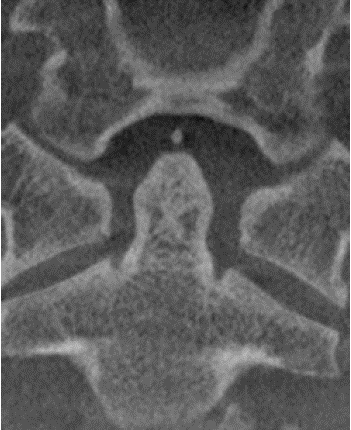
Figure 1: CBCT coronal reconstruction image of an OTP.
OO: The os odontoideum represents the separation of the odontoid tip from the body of C2, with a smooth and separate caudal portion of the odontoid process [1]. (Figure 2)
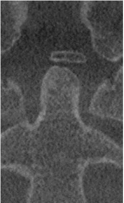
Figure 2: CBCT coronal reconstruction image of an OO.
Odontoid aplasia/hypoplasia: Excessive dysplasia that does not reach the upper edge of the anterior atlantic arch. In patients with condylar hypoplasia, the joint space is flattened, and the Atlanto-Occipital Joint Angle (AOJA) increases. AOJA measurements were performed if odontoid hypoplasia was present.
Dens bicornis: The presence of a partition from the lower synchondrosis to the tip of the odontoid process [1].
Inclination of the odontoid process: An anteverted odontoid process can bend over the anterior arch of the atlas. [1]
Malpositioning of the odontoid process: The odontoid process can be dramatically posteriorly positioned and may be located anterior to the anterior arch of the atlas [1].
Dubliorated odontoid process: Bony discontinuity of the posterior arch of C1 and a hypertrophied anterior arch. [1]
Fused nonseperated odontoid process to the anterior arch of the atlas: This defect causes abnormal placement of the ossification centers or a complete absence of the ossification center, hindering appropriate fusion of the said centers and leading to abnormal movement in the midline, creating the appearance of a fissure in the anterior arch of the atlas [1].
Dolicho-odontoid: hyperplasia and distortion of the odontoid tip with lateral or posterior deviation [1].
Only OTP and OO were detected in the evaluated tomographies. Information about the patients’ age and sex was recorded on a special form. After this, two observers (AZZ and AAP; dento-maxillofacial radiologists with 19 and 29 years of experience, respectively) independently performed the radiographic measurements.
Radiographic measurements were:
i-Dimensions of extra ossicles: Dimensions of the OTP and OO were measured in sagittal, coronal and axial images (Figure 3a and 3b).
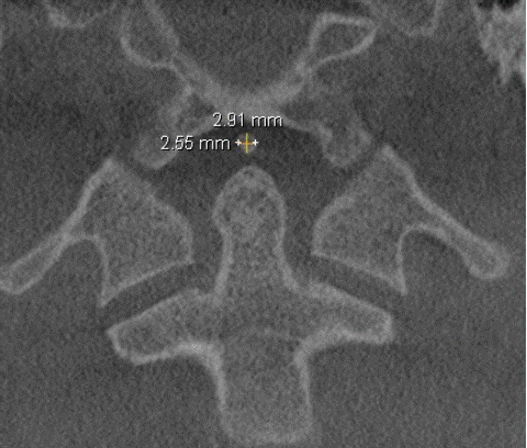
Figure 3a: CBCT coronal reconstruction image showing the dimensions of an OTP.
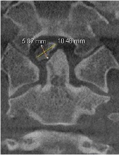
Figure 3b: CBCT coronal reconstruction image showing the dimensions of an OTP.
Measurements that were performed at the sagittal CBCT reconstruction;
ii-Extraorbital-dental interval (EDI): The distance between the inferior surface of the ossicles and the nearest surface of the odontoid process (Figure 4).
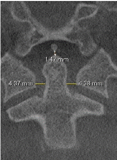
Figure 4: Cone-Beam Computed Tomography (CBCT) coronal reconstruction image showing the EDI and LADI measurements of an OTP.
iii-Anterior atlantodental interval (AADI): The distance between the odontoid process and the posterior border of the anterior arch of the atlas.
The atlantoaxial instability (AAI) was evaluated by measuring the AADI (a distance of more than 5 mm indicating atlanto-axial instability) (Figure 5).
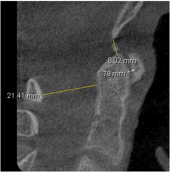
Figure 5: CBCT sagittal reconstruction image showing the measurements of the AADI, PADI and BDI of an OTP.
iv-Posterior atlantodental interval (PADI): The distance between the posterior surface of the dens and the anterior surface of the posterior arch of the atlas (Figure 5).
v-Basion-dens interval (BDI): The distance from the most inferior portion of the basion to the closest point of the superior aspect of the dens in the midsagittal plane (Figure 5).
Measurements that were performed at the coronal CBCT reconstruction;
vii- Lateral atlanto dens interval (LADI) asymmetry: the difference between left LADI and right LADI (Figure 4).
The violia-Atlanto-occipital joint angle (AOJA) was measured at the intersection of tangents drawn parallel to the atlanto-occipital joints. AOJA measurements were performed if odontoid hypoplasia was present. It is normal between 124 and 127. In condylar hypoplasia, the joint space is flattened, and the angle increases (Figure 6).
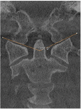
Figure 6: CBCT coronal reconstruction image showing the measurements of AOJA of an OTP.
The data were analyzed using descriptive statistics.
Results
CBCT images of fourteen patients (0.7%) showed developmental anomalies of the odontoid process. OTP was found in 10 patients, for a prevalence of 0.5%. Five of the patients with OTP were male, and five were female. The mean age of the patients was 44.46 years, ranging between 18 and 65 years. OO was observed in four patients (0.2%). All were orthotopic in type. Other developmental anomalies could not be identified.
The interobserver agreement was excellent according to kappa statistics (0.9-1.00). The sizes of the extra ossicles and the EDI, AADI, PADI, LADI asymmetry, BDI, and AOJA values for OTP and OO are presented in Table 1 and Table 2, respectively. The mean AADI was found to be 1.2 mm in males and 1.1 mm in females. In one of the OTP patients, the AOJA was greater than 127, but the length of the odontoid process was 16.9 mm. AAI was not identified among OTP or OO patients.
OTP (n)
Sex
D1
D2
D3
EDI
AADI
PADI
LADI
asymBDI
AOJA
1
E
2.6
3.0
3.2
1.5
1.8
21.4
0
6.0
123
2
E
2.0
2.3
1.8
1.7
1.8
17.4
0.5
5.3
117
3
K
1.2
1.8
1.7
1.3
1.6
14.2
0.6
4.9
117
4
E
3.3
2.4
4.3
1.3
1.7
16.2
0.6
4.8
135
5
E
3.3
2.8
4.0
0.1
1.6
17.6
1.3
8.4
112
6
K
5.1
3.3
3.8
0.4
1.2
19.2
0.7
5.1
115
7
K
1.7
1.5
1.9
1.3
0.5
21.0
0.5
4.1
110
8
E
3.1
1.5
3.8
0.5
1.2
22.1
0.3
4.6
121
9
E
2.4
2.6
2.3
0.2
0.5
22.0
2.0
3.6
105
10
E
3.4
2.9
3.6
0
0.5
20.7
0.1
4.4
103
OTP: Ossiculum Terminale Persistans. D1: Horizontal Dimension. D2: Vertical Dimension. D3: Sagittal Dimension; EDI: Extraossicle-Dental Interval. AADI: Anterior Atlantodental Interval; PADI: Posterior Atlantodental Interval; LADI asym: Lateral Atlanto Dens Interval Asymmetry. BDI: Basion-Dens Interval. AOJA: Atlanto-Occipital Joint Angle
*Measurements are in millimeters
Table 1: Size of OTPs and radiographic measurements.
OO (n)
Sex
D1
D2
D3
EDI
AADI
PADI
LADI
asymBDI
AOJA
1
K
10.5
5.9
4.7
2.0
1.9
16.4
0.9
5.2
115
2
E
8.9
3.6
4.7
0.8
1.2
19.2
1.4
8.5
106
3
K
2.4
2.9
2.5
1.0
1.4
19.2
1.3
2.7
129
4
K
8.4
3.7
3.8
0.7
0.4
17.2
0.2
4.2
116
OO: Os Odontoideum. D1: Horizontal Dimension. D2: Vertical Dimension. D3: Sagittal Dimension; EDI: Extraossicle-Dental Interval; AADI: Anterior Atlantodental Interval; PADI: Posterior Atlantodental Interval; LADI asym: Lateral Atlanto Dens Interval Asymmetry; BDI: Basion-Dens Interval; AOJA: Atlanto-Occipital Joint Angle
*Measurements are in millimeters
Table 2: OOs size and radiographic measurements.
Discussion
The odontoid process has been observed to have many congenital and acquired anatomical variants. Congenital anomalies include various types of odontoid dysgenesis, such as os odontoideum, persistent os-terminale, odontoid aplasia or hypoplasia, dens bicornis, inclination of the odontoid process, malposition of the odontoid process, dublicated odontoid process, fused nonseperated odontoid process to the anterior arch of the atlas, and dolicho-odontoid [1]. Acquired anomalies of the odontoid may be traumatic, degenerative, inflammatory, or neoplastic. The etiology of this disease is not fully understood but is likely multifactorial.
The prevalence [13] of congenital cervical vertebra anomalies has been reported to range from 0.9% to 41.5%; a greater number of congenital cervical vertebra anomalies is likely based on false-positive findings on lateral cephalometric radiographs [15]. Alsufyani NA [13]. evaluated the incidental findings in the cervical vertebra and clivus on CBCT images and reported that 16.2% of the findings were congenital. In this study, CBCT images of fourteen patients (0.7%) showed developmental anomalies of the odontoid process.
During normal development of the C2 vertebra, the odontoid process itself is derived from two lateral primary ossification centers, and the apex is derived from an apical secondary ossification center. This secondary ossification center, referred to as the ossiculum terminale, does not appear until 3 to 6 years of age and does not complete fusion with the remainder of the odontoid until the beginning of puberty [1]. However, small, well-corticated ossicles in the vicinity of C1 and in the dens of C2 fail to fuse and form OTP. The presence of OTP should not be considered pathological until it persists beyond the age at which skeletal maturity is attained [3]. Because of this information, this study was conducted with patients older than 12 years.
An increased incidence of odontoid ossicles has been reported in patients suffering from Down syndrome. Viswanathan et al. [4] reported the case of a 10-year-old boy without any syndrome or other associated abnormalities who presented with multiple falling episodes and who was found to have undiagnosed OTP on CT scan. Constantoyannis et al. [5] presented the case of a 36-year-old woman with gradual onset of neck pain and neurological signs who was also found to have OTP close to the arc of the atlas. Alsufyani NA. [13] found 14 OTP among 7689 CBCT images, 1.9% of which were incidental findings. In this study, OTP was found in ten (0.5%) patients without any syndrome or other associated abnormalities.
Os odontoideum is an uncommon diagnosis. An oval-shaped, well-corticated bony ossicle is positioned cephalad to the body of the axis. However, the etiology/pathogenesis of this lesion remains controversial. However, there are two theories about the origin of os odontoideum: traumatic or congenital. [16] The most widely accepted etiology of OO is fracture of the dens, which is often unnoticed by the patient, followed by nonunion. Conversely, the incidence of this anomaly in identical twins and people with familial relations also weakens the prior injury argument. In addition, OO has also been described in association with many other syndromes, with Down syndrome being the most common syndrome. They may be present in the normal position of the odontoid process (orthotopic), displaced cranially (dystopic), or fused to the clivus.
The most common presenting symptom associated with OO was neck pain, followed by weakness and paresthesia of the upper and lower limbs, with the upper limbs more commonly involved. Gait disturbance, neck stiffness, and headaches were uncommon presenting complaints, as was torticollis. A mixture of upper and lower motor neuron signs was present in a proportion of these patients. Minor trauma in these patients might cause sudden death [8].
Reports of OO in the dental literature are limited to a few studies [13,18]. Alsufyani NA. [13] reported only one case of OO (0.01%) as an incidental finding of the cervical spine and clivus in a retrospective CBCT study. In this study, OO was observed in four (0.2%) patients, and all the patients were in the orthotopic position. However, we do not know whether these patients have any neurologic symptoms due to OO.
The presence of OTP and OO may be clinically difficult to distinguish from a type I odontoid fracture that occurs at the tip of the dens or a type II odontoid fracture that occurs at the base of the dens. [19] Acute fractures show irregular, lucent margins on imaging, whereas OTP and OO patients present with smooth, corticated margins. Chronic type I fractures of the odontoid may exhibit nonunion and display corticated margins on imaging. In addition, ossicles must be differentiated from “joint mice” (those with loose intra-articular bodies from those with degenerative joint diseases). [13] Additionally, OTP must be differentiated from OO. Constantoyannis et al. [5] noted the difficulty of distinguishing between OTP and other odontoid anomalies, such as OO, and concluded that surgical intervention is necessary regardless of the final diagnosis if a patient is presenting with neurological signs.
One of the limitations of this study is that we could not find any other developmental or congenital anatomical variants of the odontoid process except OTP or OO among the 1950 CBCT images, and we cannot discuss their prevalence or any other features.
Atlantoaxial Instability (AAI) with normal vertebral morphology is usually evaluated by measuring the Atlas-Dens/odontoid Interval (ADI). The ADI of the atlantoaxial joint, including the anterior Atlantodental Interval (AADI) and the Lateral Atlantodental Interval (LADI), has been studied extensively using conventional radiographs and is widely used in the evaluation of AAI.
The AADI is known as the predental space. Chen et al. [20] reported that the AADI was <3.0 mm in most males (97.9%) and <2.5 mm in most females (97.7%). Wang et al. [10] reported 24 patients with OO and used 5 mm to diagnose AAI. In this study, a distance greater than 3 mm was considered to indicate AAI because of the studied adult population, and we could not detect any AAI in OTP or OO patients. The maximum AADI was 1.9 mm, which was considered the normal interspace.
Chen et al. [20] reported that the AADI was 1.83 ± 0.46 mm (0.9–3.4 mm) in males and 1.63 ± 0.43 mm (0.5–3.2 mm) in females, which indicated that the AADI was significantly greater in males than in females. In this study, the mean AADI was found to be 1.2 mm in males and 1.1 mm in females.
The LADI is routinely evaluated for the spread of lateral masses in C1 and for LADI asymmetry. Asymmetry of the LADI in patients with a history of recent trauma is indicative of rotatory subluxation of the atlas on the axis. Persistent LADI asymmetry not corrected by rotation should be used as the basic radiological criterion for the diagnosis of atlantoaxial rotatory fixation. Ajmal and O’Rourke [21] studied 13 neck injury patients with an LADI asymmetry =2.0 mm and suggested that LADI asymmetry might be a sign of significant cervical trauma. In this study, all the CBCT scans were performed with the patient in a normal head position, aiming to diminish the influence of head rotation on the results. We identified only an OTP patient with LADS asymmetry equal to 2 mm.
PADI measurements are important if they are less than 14 mm, which is associated with an increased risk of neurological injury and is an indication for surgery. [8] In this study, the smallest PADI was 14.5 mm, which was obtained for an OTP patient.
One of the measurements used in this study was the Basion-Dens Interval (BDI). At the beginning of this study, we hypothesized that the BDI would increase among OTP and OO patients because of the additional ossicles at the tip of the dens. However, the maximum BDI measurement found in an OO patient was 8.5 mm, which was within the normal limit.
Adult patients with AAI associated with OTP [4,5] and OO [10,11] have been described in the literature. An increased incidence of OTP has been reported in patients suffering from Down syndrome, and its presence may contribute to the AAI, which is often associated with this condition. In this study, the patients did not suffer from acute trauma or any syndrome, but the limitation of this retrospective study is that we cannot determine the clinical findings of these patients.
The AOJA is formed at the intersection of tangents drawn parallel to the atlanto-occipital joints. It is normal for the peak to be between 124 and 127°. In condylar hypoplasia, the joint space is flattened, and the angle increases. In this study, in one of the OTP patients, the AOJA angle was greater than 127°, but the length of the odontoid process was 16.9 mm, which was within the normal range.
Early and accurate diagnosis is critical for mitigating morbidity and mortality due to these anomalies. Minor trauma in patients with undiagnosed cervical instability might result in catastrophic neurological results [16]. Imaging has an important role in the management of developmental anomalies of the odontoid process, from diagnosis to therapy. A multimodal imaging approach, including plain radiography, CT and MRI, may be used to identify and provide the detailed anatomy of these anomalies, changes in the surrounding structure, atlanto-axial instability, spinal cord compression, associated craniocervical junction malformation and favorable anatomy to guide surgical therapy [10]. Routine anteroposterior and lateral cervical spine radiographs with an open-mouth odontoid are the first approach to this condition and can aid in diagnosis. Computed tomography images confirmed the presence of a corticated margin in this ossicle. It shows a shortened odontoid process and a smooth ossicle of bone [16,17]. Several authors also perform bedside lateral radiographs during preoperative skull traction because they consider them to be very important for atlantoaxial joint instability and subluxation and could help determine the surgical method used. [22] Multidetector CT with isotropic resolution and multiplanar reformations has enabled better visualization of complex bony abnormalities. It also helps with craniometric measurements that cannot be accounted for by plain radiographs [23].
Cone-beam images are distinguished from CT images in part by their imaging geometry. The X-ray beam in a cone-beam unit diverges as a cone to the patient rather than being collimated into a fan beam, as in a CT machine. This allows the region of interest to be imaged with one cycle of the X-ray source, or less. It is possible to extract axial, sagittal or coronal images, as well as planar or curved reconstructions on the same window. Three-dimensional (3-D) images of bone or air/soft tissue interfaces can also be generated [24]. In this study, CBCT imaging allowed us to visualize cervical vertebra and developmental anomalies of the odonoid process in three planes in one window.
Although the region of immediate diagnostic interest may be limited to a specific site, it is important to evaluate all the structures imaged. Disease is often an incidental finding that is typically located in part of the image and is unrelated to the reason for which the image was obtained. Accordingly, it is critical to systematically evaluate the entire image or imaged volume. With the expanded use of CBCT equipment in the dental profession, identification, interpretation, and management of incidental findings are necessary, as some findings require a higher level of expertise and additional advanced imaging modalities. [24] In regard to incidental findings in the cervical spine, there is scant information in the dental literature, and the pressure on OMRs is great because of the complex anatomy, complicated pathophysiology, and lack of soft tissue depiction via CBCT [13]. Early recognition and proper identification of radiological and clinical signs of AAI may help to avoid catastrophic complications secondary to spinal cord compression. Once the physician has confirmed the diagnosis, patients and their families require education from surgeons and orthopedic nurses about this condition and its possible outcomes [8]. Surgery is usually indicated for patients who have one of the following conditions: neurological involvement, radiological signs of instability, progressive instability, or persistent neck pain associated with atlantoaxial instability [9]. Conservative treatment is an option only when the patient is asymptomatic and when there is no evidence of atlantoaxial instability. Patients should be aware of the possible catastrophic consequences of this pathology [16].
Conclusion
In conclusion, within the limitations of this study, developmental anomalies of the odontoid process, such as OT and OO, are rare anomalies of the odontoid process that may cause AAI. These lesions may appear incidentally on large-FOV CBCT images. Dentomaxillofacial radiologists should be able to identify these anomalies, especially for AAI, via CBCT and note them in their reports. Awareness of these findings will be quite helpful in assessing the risk of complications and in informing patients about their condition. Awareness can also help clinicians when it is necessary to refer patients to an expert physician.
Author Statements
Funding
Authors declare that there is no source of funding or financial interest.
Conflict of Interest
Authors declare that there is no conflict of interest.
Authors Contributions
AZZ: Data collection, Data analysis, Manuscript writing/editing. Concept and design of study or acquisition of data or analysis and interpretation of data;
APS: Data analysis, Manuscript writing/editing. Drafting the article or revising it critically for important intellectual content and Final approval of the version to be published.
KC: Manuscript writing, Taking ethic permision Drafting the article or revising it critically for important intellectual content and Final approval of the version to be published.
Ethics: This study was reviewed and approved by the Institutional Review Board of the University (OMU KAEK 2022/499). All procedures followed were in accordance with the ethical standards of the responsible committee on human experimentation (institutional and national) and with the Helsinki Declaration of 1975, as revised in 2008 (5).
References
- Akobo S, Rizk E, Loukas M, Chapman JR, Oskouian RJ, Tubbs RS. The odontoid process: a comprehensive review of its anatomy, embryology, and variations. Childs Nerv Syst. 2015; 31: 2025–34.
- O’Toole P, Tomlinson L, Dormans JP. Congenital Anomalies of the Pediatric Cervical Spine. Seminars in Spine Surgery. 2011; 23: 199-205.
- Johal J, Loukas M, Fisahn C, Oskouian RJ, Tubbs RS. Bergmann’s ossicle (ossiculum terminale persistens): a brief review and differentiation from other findings of the odontoid process. Childs Nerv Syst. 2016; 32: 1603-1606.
- Viswanathan A, Whitehead WE, Luerssen TG, Illner A, Jea A. Orthotopic ossiculum terminale persistens and atlantoaxial instability in a child less than 12 years of age: a case report and review of the literature. Cases J. 2009; 2: 8530.
- Constantoyannis C, Konstantinou D, Maraziotis T, Dimopoulos PA. Atlantoaxial instability and myelopathy due to an ossiculum terminale persistens. Med Sci Monit. 2004; 10: CS63–67.
- Klassov Y, Benkovich V, Kramer MM. Posttraumatic os odontoideum - case presentation and literature review. Trauma Case Rep. 2018; 18: 46-51.
- Hedequist DJ, Mo AZ. Os Odontoideum in Children. J Am Acad Orthop Surg. 2020; 28: e100-e107.
- Pereira Duarte M, M Das J, Camino Willhuber GO. Os Odontoideum. In: StatPearls. Treasure Island (FL): StatPearls Publishing. 2022.
- Pluemvitayaporn T, Kunakornsawat S, Piyaskulkaew C, Pruttikul P, Pongpinyopap W. Chronic posterior atlantoaxial subluxation associated with os odontoideum: a rare condition. A case report and literature review. Spinal Cord Ser Cases. 2018; 4: 110.
- Wang Q, Dong S, Wang F. Os odontoideum: diagnosis and role of imaging. Surg Radiol Anat. 2020; 42: 155-160.
- Dias RP, Buchanan CR, Thomas N, Lim S, Solanki G, Connor SEJ, et al. Os odontoideum in Wolcott-Rallison syndrome: a case series of 4 patients. Orphanet J Rare Dis. 2016; 1: 1–5.
- Sanjay M. Mallya. Radiographic Anatomy. In: Sanjay M. Mallya and Ernest W.N. Lam (eds). White and Pharoah’s Oral Radiology. Principles and Interpretation. Oral radiology principles and interpretation, 8th ed. Missouri: Elsevier Mosby, 2019.
- Alsufyani NA. Cone beam computed tomography incidental findings of the cervical spine and clivus: retrospective analysis and review of the literature. Oral Surg Oral Med Oral Pathol Oral Radiol. 2017; 123: e197-e217.
- Raj A, Srivastava SK, Marathe N, Bhosale S, Purohit S. Dystopic Os Odontoideum Causing Cervical Myelopathy: A Rare Case Report and Review of Literature. Asian J Neurosurg. 2020; 15: 236-240.
- Bebnowski D, Hänggi MP, Markic G, Roos M, Peltomäki T. Cervical vertebrae anomalies in subjects with class II malocclusion assessed by lateral cephalogram and cone beam computed tomography. Eur J Orthod. 2012; 34: 226-31.
- Duarte MP, Das JM, Willhuber GOC. Os Odontoideum. StatPearls publishing; 2024-2023.
- Tang X, Tan M, Yi P, Yang F, Hao Q. Atlantoaxial dislocation and os odontoideum in two identical twins: perspectives on etiology. Eur Spine J. 2018; 27: 259-263.
- Tetradis S, Kantor ML. Anomalies of the odontoid process discovered as incidental findings on cephalometric radiographs. Am J Orthod Dentofac Orthop. 2003; 124: 184-189.
- Goel A, Cacciola F. The craniovertebebral junction: diagnosis, pathology, surgical techniques. Thieme, Stuttgart. 2011.
- Chen Y, Zhuang Z, Qi W, Yang H, Chen Z, Wang X, et al. A three-dimensional study of the atlantodental interval in a normal Chinese population using reformatted computed tomography. Surg Radiol Anat. 2011; 33: 801-6.
- Ajmal M, O’Rourke SK. Odontoid Lateral Mass Interval (OLMI) asymmetry and rotary subluxation: a retrospective study in cervical spine injury. J Surg Orthop Adv. 2005; 14: 23–26.
- Wu X, Wood KB, Gao Y, Li S, Wang J, Ge T, et al. Surgical strategies for the treatment of os odontoideum with atlantoaxial dislocation. J Neurosurg Spine. 2018; 28: 131-139.
- Jain N, Verma R, Garga UC, Baruah BP, Jain SK, Bhaskar SN. CT and MR imaging of odontoid abnormalities: A pictorial review. Indian J Radiol Imaging. 2016; 26: 108-19.
- Scarfe WC. Incidental findings on cone beam computed tomographic images: a Pandora’s box? Oral Surg Oral Med Oral Pathol Oral Radiol. 2014; 117: 537-540.