Clinical Image
Austin J Radiol. 2025; 12(3): 1259.
Hairy Kidney Sign a Must Know Diagnosis
Soukayna J*, Khadija ElA, Jamal ElF and Tarik S
Radiology Department, National Institute of Oncology, Rabat, Morocco
*Corresponding author: Jabour Soukayna, Radiology Department, National Institute of Oncology, Rabat, Morocco Email: dr.jaboursoukayna@gmail.com
Received: July 14, 2025 Accepted: July 24, 2025 Published: July 25, 2025
Clinical Image
Keywords: Rare disease; CT imaging; Early diagnosis
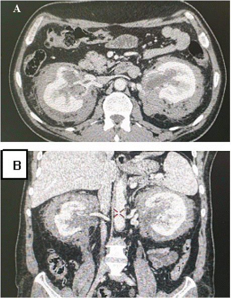
Discussion
Erdheim-Chester disease (ECD) is a rare, acquired form of non- Langerhans cell histiocytosis affecting adults. It is characterized by xanthogranulomatous infiltration of lipid-laden histiocytes and presents with a wide range of systemic symptoms. The most common clinical features involve the skeleton—particularly the long bones—which often show increased uptake on technetium-99m bone scintigraphy [1], and the urinary tract, notably with retroperitoneal fibrotic changes. Diagnosis is typically confirmed through ultrasoundguided biopsy of the perirenal infiltrative tissue.
Urological manifestations are present in approximately 30% of ECD cases. These frequently appear as retroperitoneal fibrosis, which can progress to bilateral hydronephrosis and impaired renal function. In contrast to idiopathic retroperitoneal fibrosis (RPF), ECDassociated fibrosis tends to spare the ureters and does not involve the inferior vena cava. A distinguishing feature is the circumferential encasement of the aorta, whereas idiopathic RPF usually preserves the posterior aspect of the vessel. ECD should be considered in all patients presenting with corticosteroid-resistant retroperitoneal fibrosis. Special attention should be paid to the imaging appearance of the perirenal fascia and fat, which may exhibit the characteristic perirenal rind or "hairy kidney" sign (Figure 1).