
Clinical Image
Austin J Radiol. 2020; 7(1): 1105.
A Rare Case of Cutaneous Mucormycosis in a Person with No Predisposing Factors
Kalani SA¹* and Shah CH²
¹Department of Medicine, Royal Blackburn Hospital, UK
²Department of Radiology, UCMS, India
*Corresponding author: Shitanshu Kalani, Department of Medicine, Royal Blackburn Hospital, UK
Received: January 27, 2020; Accepted: January 29, 2020; Published: February 05, 2020
Clinical Image
A 57-year-old male presented with painful, purulent swelling on the posterior medial aspect of the right ankle joint (Figure 1). The patient mentioned this had been getting worse since the last 1 week but didn’t recall when it actually started. A detailed history was taken and there was no history of trauma, fever or systemic symptoms. The patient had no history of diabetes or immunosuppression. An X ray of the right ankle showed a mass posterior to calcaneum (Figures 2 and 3). Subsequent MRI report mentioned that the lesion showed haemorrhagic and cystic areas with increased vascularity and surrounding soft tissue edema. The lesion was biopsied and the findings were in keeping with a diagnosis of cutaneous invasive mucormycosis.
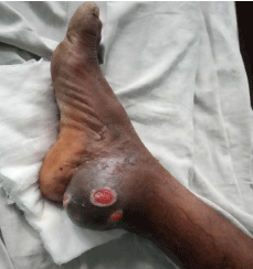
    Figure 1: Clinical picture; a large mass can be seen on the posterior medial
aspect of right ankle joint with definite changes on the overlying skin, raising
high suspicion for malignancy at the first glance.
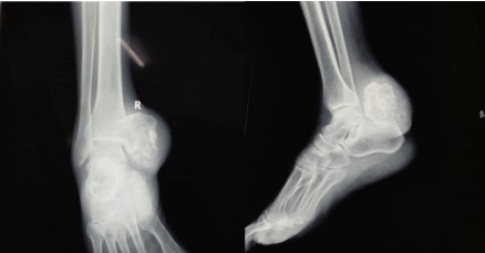
    Figure 2&3: It can be seen that there is increase in soft tissue in retrocalcaneal
location causing obstruction of Kaeger’s fat pad extending to the medial
aspect of knee joint. Dense central calcification seen within the soft tissue.
The calcification laterally is in close contact with the medial malleolus. Rest of
the bones and joints are normal.
Rhinocerebral/rhinofacial form is the most common but occasionally pulmonary, gastrointestinal and cutaneous forms may also occur. Mucormycosis of the cutaneous and subcutaneous tissues is relatively uncommon. Mortality varies depending on site of infection and patient co-morbidity. Single site infection in patients with no underlying pathology has an overall mortality of 35% but disseminated disease has a mortality of 96%. The most common predisposing factors include Diabetes, immunosuppression and history of trauma. Patients with chronic renal failure are also susceptible. Mucormycosis can be diagnosed in histopathologic sections by Hematoxylin and Eosin stain (H&E) with the aid of PAS or GMS stain. Microscopically, the fungus is characterized by broad, non-septate hyphae that are either cylindrical, irregular or distorted in shape. The hyphal branching is irregular and the walls of the hyphae vary in thickness. The fungal infection is almost always accompanied by either a granulomatous infiltrate composed of epithelioid and multinucleate giant cells or a polymorphonuclear leukocyte infiltrate forming micro abscesses. Routine blood work may not suggest anything and the cultures may not grow any organisms.