Case Presentation
Austin J Radiol. 2020; 7(2): 1110.
Unusual Combination Hypertrophic Cardiomyopathy and Arrhythmogenic Cardiomyopathy Phenotype
Mashego B and Nethononda MR*
Division of Cardiology, University of the Witwatersrand, South Africa
*Corresponding author: Nethononda MR, Division of Cardiology, Chris Hani Baragwanath Academic Hospital and the University of the Witwatersrand, PO Bersham, 2013, Johannesburg, South Africa
Received: May 27, 2020; Accepted: June 25, 2020; Published: July 02, 2020
Abstract
Inherited forms of cardiomyopathy such as hypertrophic as well as arrthymogenic right ventricular cardiomyopathies are amongst the commonest cause of sudden cardiac death particularly in young people. Although these conditions both have an autosomal dominant mode of transmission, the genetic mutations and affected proteins are different. We present a unique case of phenotypic combination of ARVC in the right ventricle and HCM on the left in a 22 years old obese female who presented with heart failure at 20 weeks of gestation. Her 12-lead resting ECG showed epsilon wave in one of the limb leads. Her Cardiac MRI showed multiple aneurysms in the right ventricular free wall suggestive of ARVC, whereas she had significant asymmetric segmental left ventricular hypertrophy and associated diffuse late gadolinium enhancement typical of hypertrophic cardiomyopathy. Her initial 24 ambulatory monitoring revealed no arrhythmias. Her heart failure was treated with beta blocker and diuretic, and she went on to deliver a healthy baby at term. Although unusual, this case demonstrates that this potential lethal combination can occur, and this possibility should be investigated with the novel technique of cardiac MRI.
Keywords: Arrhythmogenic ventricular cardiomyopathy; Hypertrophic cardiomyopathy; Cardiac MRI
Case Presentation
A 22 years old female presented to our Hospital with progressive shortness of breath and leg edema at 20 weeks of gestation with her first pregnancy. She had no prior history of hypertension or other chronic conditions. On physical examination she was obese with a body mass index of 38.1kg/m2 and a body surface area of 1.95m²; a regular pulse of 90bpm; blood pressure of 120/67mmHg, respiratory rate 22breath/min; O2 saturation 95%; Jugular venous pressure was not raised; there was a 2/6 pansystolic murmur at the base; there were a few basal crepitation in both lungs. She was treated with diuretics and beta blocker. The course of her pregnancy was uneventful, and she delivered a healthy baby at term.
Resting ECG revealed sinus rhythm, bi-phasic P wave in V1, leftward QRS axis shift and epsilon waves in lead aVL (Figure 1). 24 hour ambulatory ECG monitoring showed no evidence of atrial fibrillation or ventricular tachycardia. Trans-Thoracic Echocardiography (TTE) Showed Asymmetric Septal Hypertrophy (ASH), diastolic left ventricular dysfunction, left atrial enlargement and right ventricular free-wall suspicious of wall motion abnormalities.

Figure 1: Blue arrow indicates a post-excitation or an epsilon wave.
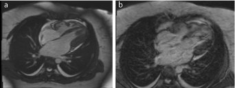
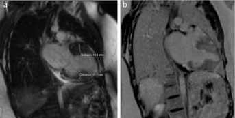
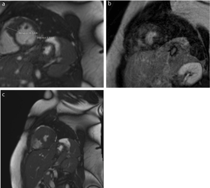
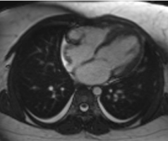
On cardiac MRI, the left ventricular mass indexed for the BSA was 64g/m2, which is towards the upper range of normal for Caucasian females (31–79g/m²). The Left Ventricular Ejection Fraction (LVEF) was 82%, also at the upper range of normal (57-81%). The Right Ventricular End-Diastolic Volume Indexed (RVEDVI) for BSA was 67ml/m², with an ejection fraction of 59%. However, there were significant structural abnormalities consistent with both hypertrophic cardiomyopathy in the LV and arrhythmogenic ventricular dysplasia in the RV as shown in the Figures (Figures 2,3,4,5).

    Figure 2: CMR end-diastolic still frame, Late Gadolinium Enhancement
(LGE) and cine horizontal long axis (HLA) images of the heart showing
aneurysmal right ventricular free wall as well as hypertrophic inferolateral wall
of the left ventricle and prominent papillary muscle (a) together with hyperenhancement
on LGE imaging (b).

    Figure 3: CMR end-diastolic still frames vertical long axis (VLA) images of
the left ventricle showing hypertrophy of the basal anterior (19mm) and midinferior
(15mm) (a) wall as well as hyper enhancement of those segments on
LGE imaging (b).

    Figure 4: (a) show end-diastolic still frames of mid-ventricular Short Axis
(SA) images of the left and right ventricle showing focal hypertrophy of the
antero-septal (29mm) segments as well as hyper enhancement of those
segments on LGE imaging (b). Figure 4 (c) in the end-systolic frame of 4 (a).

    Figure 5: CMR end-diastolic still frame and cine transverse images of the
heart showing aneurysmal right ventricular free wall as well as hypertrophy of
the mid to apical septum.
Discussion
Both ARVC and HCM are inherited forms of cardiomyopathy, usually autosomal-dominant that are associated with sudden cardiac death particularly in young people [1]. The prevalence of ARVC is 1:2000 but is higher in certain regions of the world such as the Greek island of Naxos [2]. On the contrary HCM is a fairly common condition with a population prevalence of 0.02 to 0.2%. We are not aware of any previous reports of a case with combined phenotypic features of ARCV and HCM. Therefore, our patient is unique in that respect. Right ventricular involvement in patients with HCM is usually characterized by hypertrophy of various regions of the RV with aneurysmal right ventricular aneurysm described in one case only as a results of localized RV infarction [3]. This is not the case in our patient who has ECG features of ARVC and extensive aneurysms of the RV free wall on CMR imaging. Her right ventricular volume indexed for her BSA on CMR was below the cut-off value of 100ml/m2, stipulated by the Task Force Criteria [4]. We believe this to be due to her younger age of 22 at presentation and subsequent CMR imaging in a few years’ time may demonstrate increasing RV volumes. Indeed, she appears to have an aggressive disease process with extensive late gadolinium enhancement. This and the myocardial wall thickness of up to 29mm in some segments are risk markers for sudden cardiac death [5]. Currently, she is not keen on ICD. She presented with heart failure that was most likely precipitated by the stress of pregnancy and will need to be watched closely for dilatation of the right ventricle and development of arrhythmias.
Conclusion
Our patient is unusual in a number of aspects; firstly, she presented for the first time in pregnancy with cardiac morphological features of both ARVC and HCM, a double wormy as both are the commonest cause of sudden cardiac death amongst young people; and secondly at age 22 she already has risk markers for sudden cardiac death. Due to limited ability for genotyping we have thus far not performed any genotyping, which will have critical implications for her family members including her newborn 1st child.
References
- Hensley N, Dietrich J, Nyhan D, Mitter N, Yee MS, Brady M. Hypertrophic cardiomyopathy: a review. Anesthesia and analgesia. 2015; 120: 554-569.
- Saguner AM, Brunckhorst C, Duru F. Arrhythmogenic ventricular cardiomyopathy: A paradigm shift from right to biventricular disease. World journal of cardiology. 2014; 6: 154-174.
- Mozaffarian D, Caldwell JH. Right ventricular involvement in hypertrophic cardiomyopathy: a case report and literature review. Clinical cardiology. 2001; 24: 2-8.
- Marcus FI, McKenna WJ, Sherrill D, Basso C, Bauce B, Bluemke DA, et al. Diagnosis of arrhythmogenic right ventricular cardiomyopathy/dysplasia: proposed modification of the Task Force Criteria. Eur Heart J. 2010; 31: 806- 814.
- Tower-Rader A, Kramer CM, Neubauer S, Nagueh SF, Desai MY. Multimodality Imaging in Hypertrophic Cardiomyopathy for Risk Stratification. Circulation Cardiovascular imaging. 2020; 13: e009026.