Clinical Image
Austin J Radiol. 2020; 7(2): 1112.
Liver Scalloping : An Evocative Sign of Pseudomyxoma Peritonei
Taibi B*, Omor O and Latib R
Department of Radiology, Mohammed V University, Morroco
*Corresponding author: Taibi B, Department of Radiology, Mohammed V University, National Institute of Oncology, Rabat Morroco
Received: June 22, 2020; Accepted: June 29, 2020; Published: July 06, 2020
Clinical Image
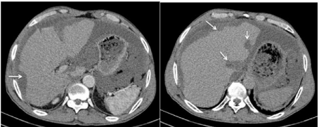
A 62 year old male presented with abdominal enlargement and pain, since 3 months. With a past history of an appendicectomy 5 years ago for a perforated appendix. Images from the ct scann revealed liver scalloping due to extrinsic compression of the liver by the gelatinous mass, partitioning, and the peritoneal effusion. It is noteworthy that this scalloping was observed even though the excreted volume was not very high. Exploratory laparotomy revealed gelatinous material in the peritoneum with seeding into the omentum.
Pseudomyxoma Peritonei (PMP), is a rare disease. It is characterized by the presence of a large amount of mucin in the abdomen [1].
If the CT aspect is similar to that observed in peritoneal carcinosis [2], there are, however, radiological semiological elements suggestive of a pseudomyxoma which are an important liver “scalloping” by the gelatinous masses, the compartmentalization of the peritoneal effusion and the presence of curvilinear calcifications (Figure 1) [3].

    Figure 1: Abdominal CT in axial section after injection of contrast agent
showing a well-designed liver “scalloping” (arrows) exerted by a peritoneal pseudomyxoma of appendicular origin.
References
- Gillion JF, Franco D, Chapuis O, Serpeau D, Convard JP, Jullès MC, et al. Appendiceal mucoceles, pseudomyxoma peritonei and appendiceal mucinous neoplasms: update on the contribution of imaging to choice of surgical approach. J Chir (Paris). 2009; 146: 150-166.
- Esquivel J, Sugarbaker PH. Clinical presentation of pseudomyxoma peritonei syndrome. Br J Surg. 2000; 87: 1414-1418.
- Diop AD, Fontarensky M, Montoriol PF, Ines DD. CT imaging of peritoneal carcinomatosis and its mimics. Diagn Interv Imaging. 2014; 95: 861-872.