Clinical Image
Austin J Radiol. 2020; 7(3): 1114.
Aortocaval Fistula
Figl J1*, Papeš D1 and Dobrota S2
¹Department of Surgery, University of Zagreb, Croatia
²Department of Radiology, University of Zagreb, Croatia
*Corresponding author: Figl J, Department of Surgery, University of Zagreb, Kišpaticeva 12, 10000 Zagreb, Croatia
Received: July 15, 2020; Accepted: July 21, 2020; Published: July 28, 2020
Clinical Image
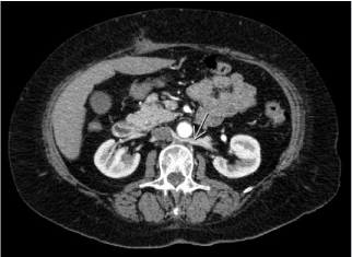
A 69-year-old male was brought to neurology emergency department due to four-hour-lasting leg-numbness and swelling. Further physical examination revealed absolutely painless pulsatile abdominal mass with only hypotension present – 95/75 mmHg and slightly troublesome breathing. Computed Tomographic Angiography (CTA) showed a previously unknown, 10-cmdiametered, juxtarenal Abdominal Aortic Aneurysm (AAA) with an extremely rare form of its rupture – a painless Aortocaval Fistula (ACF) and pulmonary embolism (Figures 1 and 2). The ACF, although very rare entity, completely explains a hypotension (ruptured AAA), a painless abdomen and patient’s good general condition despite ruptured AAA – owing it to lack of retroperitoneal distension and also breathing-difficulties due to paradox pulmonary embolism and leg-swelling and numbness (suddenly increased venous flow).

    Figure 1: Arterial phase of MSCTA with contrasting media simultaneously in
both aorta, iliac veins and inferior cava vein – a pathognomonic sign of ACF.
