Research Article
Austin J Radiol. 2021; 8(1): 1119.
The Ground-Glass Ethmoid Sinus Sign" May be the Sole Indicator for Acute Sinusitis on CT: A New Sign at Paranasal Sinus CT Images
Duzgun Yildirim1*, Deniz Esin Tekcan2, Özlem Akinci3, Elvan Cevizci AkkiliÇ, Aytug Altundag5
1Department of Medical Imaging, Acibadem University, Vocational School of Health Sciences, Istanbul, Turkey
2Department of Radiology, Acibadem University, Istanbul, Turkey
3Department of Radiology, Sancaktepe Professor Ilhan Varank Training and Research Hospital, Istanbul, Turkey
4Department of Neurology, Acibadem University, Istanbul, Turkey
5Department of Otorhinolaryngology, Biruni university, Istanbul, Turkey
*Corresponding author: Yildirim D, Department of Radiology, Acibadem University, Inonu St. No:24, Kozyatagi, 34734, Istanbul, Turkey
Received: December 22, 2020; Accepted: January 12, 2021; Published: January 19, 2021
Abstract
Objective: In this retrospective study we aimed to investigate the diagnostic value of “Ground-Glass Ethmoid Sinus Sign” (GGESS) in ethmoid cells in patients with clinical acute sinusitis.
Patients and Methods: Between January 2018-December 2018, 440 CT of the paranasal region taken for any reason in our clinic were evaluated. Mucosal thickening of the paranasal sinus wall, secretion levels, secretion bands, and Ground-Glass Signs on Ethmoid cellular walls (GGESS) were evaluated by two radiologists. The diagnostic significance of GGESS in patients with clinically positive findings and those without a diagnosis of sinusitis was statistically analyzed.
Results: Patients were included in the study classified as having acute sinusitis (Group 1-103 cases) and without a clinical history of acute sinusitis (Group 2-337 cases). In the diagnosis of acute sinusitis, GGESS had a positive predictive value of 79%, a negative predictive value of 95%, a sensitivity of 86%, and a specifity of 93%. The GGESS finding was found to be significantly higher in the acute sinusitis group as 86%, while it was 7% in the asymptomatic group (p<0.001).
Conclusion: The presence of “GGESS” on paranasal sinus CT images is associated with acute sinusitis significantly more than any other sinus inflammation findings.
Keywords: Acute sinusitis, Paranasal sinus CT, Ground-glass ethmoid sinus, Radiological diagnosis
Abbreviations
CT: Computed Tomography; GGESS: Ground Glass Ethmoid Sinus Sign; FESS: Functional Endoscopic Sinus Surgery; SPSS: Statistical Package for the Social Sciences; SD: Standart Deviation
Introduction
Acute sinusitis is the inflammation of one or more paranasal sinuses lasting less than four weeks. Clinically, it is characterized by nasal congestion, rhinorrhoea, purulent post-nasal / nasal discharge, facial fullness, and pain, headache, cough [1]. Although it is diagnosed usually with clinical and examination findings, imaging methods are used to exclude any underlying organic or obstructive pathology due to the persistence of the disease, prolongation of symptoms, prolonged or severe relapses, or frequent recurrence [2]. The apparent loss of aeration and significant effusion levels appear on direct radiographs, but further examination is usually required for false positive and false negative rates due to the inability to distinguish other etiologies that cause opacification, such as infection-inflammation-neoplasia, and differences in interobserver evaluation [3-5]. Because of improved visualization of sinus anatomy CT frequently used modality in the management of sinusoidal problems and the selection of medical/ surgical treatment according to the etiology [6,7]. Findings on CT suggestive of sinusitis include thickened mucosa (> 4 mm), air fluid levels, and opacification of the sinuses [8]. The literature on the conventional signs for the usefulness of CT in the diagnosis of rhinosinusitis remains a question of concern [9,10]. Usually ground glass appearance in the paranasal sinus refers to a fibrous osseous lesion [11]. However, the concept of Ground Glass Ethmoid Sinus Sign (GGESS) has never been used in the literature yet. By “GGESS” is meant ground glass observed in the ethmoid sinus without any pathology in the surrounding bone tissues. Contrary to belief, when ground-glass ethmoid sinus sign is searched, we believe that CT’s contribution to diagnosis in acute sinusitis radiological evaluation will rise to the level it deserves.
In this retrospective study, we aimed to determine the diagnostic value of GGESS, regardless of etiology (viral, bacterial) or the extent of the disease (sinusitis, rhinosinusitis) in patients with acute sinusitis clinic.
Materials and Methods
Paranasal sinus and craniomaxillofacial CT images of 440 patients admitted to our radiology unit between January 2018 and December 2018 were evaluated retrospectively.
After the paranasal sinus area was scanned with 0.625 mm collimation on a dual-source CT device with 128x2 detector, sharp edge structures were formed by B70 kernel, coronal plane bone algorithm (Siemens, Flash Definition, Erlangen, Germany); and ethmoid sinus walls were examined in multiplanar images at workstations.
CT was not performed in patients with single attack-simple acute sinusitis clinic and who responded to medical treatment. Paranasal sinus CT was performed in symptomatic patients (facial pain-fullness, purulent rhinitis, postnasal discharge, hyposmia-anosmia, cough) who had recurrent episodes of sinusitis (less than 3 within 1 year) or did not respond to empirical treatment. The patients in the control group consisted of patients who underwent craniomaxillofacial CT for any reason, with the most common indication for headaches, and whose other complaints related to acute sinusitis were unknown. Patients with chronic-complicated rhinosinusitis (sinusitis symptoms lasting more than 12 weeks, or complicated acute sinusitis) known Functional Endoscopic Sinus Surgery (FESS) and similar history of sinus surgery were not included in the study.
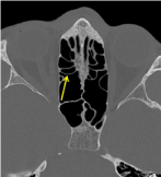
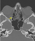
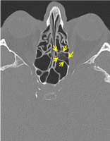
CT images of 103 patients’ who were evaluated in favor of acute sinusitis with the symptomatology and clinical examination by the clinician, and 337 patients’ who we accepted as a control group was examined by two readers (DY, experienced in head and neck radiology for 18 years and, D.E.Texperienced for 4 years). Mucosal thickening of the paranasal sinus walls (> 2 mm), effusion, and ethmoid cellular wall mucosal Ground-Glass Sign (GGESS) were recorded. Images have been reported for the consensus of this radiological imaging finding (Figure 1,2,3).

Figure 1: A normal view of paranasal ethmoid sinuses on the axial section of paranasal sinus CT.

    Figure 2: Dependent wall thickening of ethmoid cellular on the axial section
of paranasal sinus CT.

Figure 3: Typical “Ground-Glass Ethmoid Sinus Sign” on the axial section of paranasal sinus CT.
Ethics committee approval was received for this study from the local ethics committee (No: 2019-3/4). The study complied with the Declaration of Helsinki. Patients were not required to give their informed consent for inclusion in this retrospective study, because we used anonymous clinical data and individual cannot be identified according to the data present.
Statistical analysis
All the data of 440 cases were analyzed statistically by using the Chi-square test in SPSS 11 program for Windows (Chicago, IL). The data were presented as mean ±SD. Categorical variables were analyzed by using the Chi-Square test and continuous variables were analyzed by using independent samples t-test. A p<0.05 value was considered statistically significant.
Results
232 of the patients included in the study were female and 208 were male, with an average age of 37. Of 103 patients with acute sinusitis symptoms (Group-1), 61 were female and 42 were male. Of the 337 patients whose paranasal sinus sections were taken for other reasons with unknown acute sinusitis symptoms (Group-2), 147 were female and 190 were male. Mucosal thickening was detected in 66 of the 103 cases in Group-1(64%), secretion levels in 11 (10.5%), secretion bands in 22 (21%), and GGESS in 89 patients (86%) (Table 1). In the symptomatic group, only GGESS was found in 23 patients (22%) who did not have any other imaging findings of acute inflammation or sinusitis. Of the 337 cases in Group-2, 148 (44%) had mucosal thickening, 2 (0.5%) secretion levels, 17 (5%) secretion bands, 23 (7%) GGESS was detected.
    Group-1 n:103 (%)
    Group-2 n:337 (%)
    p


    Mucosal thickening
    66 (64)
    148 (44)
    <0.001


    Secretion Levels
    11 (10.5)
    2 (0.5)
    <0.001


    Secretion Bands
    22 (21)
    17 (5)
    <0.001


    Ground-Glass Ethmoid Sinus
    89 (86)
    23 (7)
    <0.001


    Chi-square test
Table 1: Comparison of CT findings between groups.
When all these data were analyzed; GGESS had a positive predictive value of 79%, a negative predictive value of 95%, a sensitivity of 86%, and a specificity of 93% in the diagnosis of acute sinusitis. The GGESS finding was found to be significantly higher in the acute sinusitis group as 86%, while it was 7% in the asymptomatic group (p<0.001).
Discussion
CT examination has an important role among all radiological methods in revealing the anatomy and abnormalities of paranasal sinuses, especially for sphenoid and ethmoid sinuses [2,12-14]. CT provides superb anatomical details and enables a fairly accurate diagnosis and delineation of the disease, addressing all concerns of the endoscopic surgeon prior to intervention [12]. The neighborhood of paranasal sinuses, especially ethmoid sinuses, to orbital and intracranial compartments makes the diagnosis and treatment of diseases in this strategic region even more important [15,16]. In untreated ethmoid sinusitis, orbital-periorbital complications may cause blindness and intracranial complications that may lead to high morbidity and mortality are very common [17,18].
Radiological imaging is not required for simple acute sinusitis attacks that are noncomplicated and respond to medical therapy. Besides, paranasal sinus CT imaging is required to evaluate the underlying organic pathologies and extent of disease infrequent attacks or refractory patients. Since it is possible to obtain important anatomical and pathological information with coronal-sagittalaxial post-processing reformats with a relatively low radiation dose equivalent to 4 plan plain radiography in new generation CT systems, it is only a matter of time to use it as first-line imaging in the diagnosis of simple/uncomplicated acute sinusitis. Air-fluid levels, mucosal thickening, and opacification of sinus cavities are considered as the characteristic findings on CT in cases with acute sinusitis [19]. Mucosal findings are nonspecific findings that all other allergic reactions, chronic events may cause mucosal thickening. Also, it may not correlate with the severity of symptoms [20,21]. Mucosal sinus findings can be detected incidentally, with rates vary between 16-40% in different patient populations [22]. In this study, the rate of mucosal thickening was determined as 64% in patients with acute sinusitis, higher than the literature [2]. However, these findings were determined with a high rate of 44% in the control group, too. Frequent and exaggerated pathological reporting of this variable degree of mucosal thickness is the cause of overdiagnosis. On the other hand, in much craniomaxillofacial imaging in daily practice, it can be reported entirely normal due to neglect of sphenoid and ethmoid sinus findings, which are highly acute and symptomatic, and exacerbation of sinusitis in these patients without treatment. Therefore, it is important to report sinusoidal findings on craniomaxillofacial CT examinations other than paranasal sinus CT.
When there is inflammation in the sinuses, the reaction that occurs primarily leads to thin mucosal thickening and effusion. This fluid may be so thin and/or also cleaned by physiologically, that it may be difficult to select it in cross-sectional images. Frequently seen mucosal thickening in the maxillary sinuses, thick osseous walls of the frontal and sphenoid sinuses or deep anatomical localizations make it difficult to evaluate this effusion. However, these thin effusions may be better visualized at the level of the ethmoid sinuses, because the thin-walled septations in the ethmoid sinuses form optimal interfaces. Therefore, it seems more logical to look for this finding in ethmoid sinuses.
Mucosal thickening alone has low sensitivity and specificity in detecting acute sinusitis. In a previous study in the literature, a high correlation was found for the diagnosis of acute sinusitis when total sinus opacification, frothy secretion and fluid levels were evaluated together. However, the positive predictive value of these symptoms alone was found to be low. In that study, for example, the positive predictive value of frothy secretion was 53% and its negative predictive value was 89% [23]. The “GGESS”, which we defined for the first time in this study, is different from the frothy secretion finding previously described in the literature and refers to the isolated ground glass image in the ethmoid sinus. In our study, a positive predictive value of GGESS was found to be 79%, a negative predictive value of 95%, and a sensitivity of 86% in the diagnosis of acute sinusitis.
The previous studies in the literature associated with acute sinusitis and radiological findings are often focused on maxillary sinus [24,25]. CT findings on ethmoid sinus were mostly emphasized in this study, and it was found that there was an increase in groundglass density in the dependent walls of the ethmoid sinus before the characteristic findings of sinusitis developed in the other sinuses. While the incidence was 7% in the control group and 86% in the acute sinusitis group, it was found to be highly positive with a significant difference. This indicates that GGESS was more valuable than conventional findings such as mucosal thickening-secretion level in the diagnosis of acute sinusitis. In our study, only GGESS was detected in 23 cases (22%) who did not have any other radiological findings of acute inflammation, such as mucosal thickening or secretion levels in the symptomatic group. It indicates that GGESS may be predictable for acute sinusitis diagnosis earlier than other findings. Thus, even if there are no other acute sinusitis findings on CT examination, reporting this finding in cases with positive clinical status will increase the diagnosis rate.
We think that GGESS, which is the sign that we consider as an early imaging finding of sinusitis, may also be seen in acute exacerbations of chronic sinusitis. The radiological findings of chronic sinusitis such as concomitant sclerotic thickened bone, intrasinusoidal calcifications, diffuse mucosal thickening are often present in this situation.
Numerous limitations were involved in this study. First, because of the limitation of radiation exposure, CT could not be applied to all patients with acute sinusitis clinic. For the same reason, the response of the imaging findings to medical treatment could not be evaluated radiologically. Another limitation of the study was the lower number of patients in the symptomatic group compared to the control group. Over time, more accurate data can be obtained by increasing the number of symptomatic cases. Although CT images were complemented similarly by secondary reconstructions on the coronal and axial plane, the different imaging protocols of the control group was another limitation of the study.
Conclusion
On paranasal sinus CT examinations; the presence of ‘’Groundglass ethmoid sinus sign’’ on the ethmoid sinus walls is associated with acute sinusitis more significantly than any other signs. Therefore, if a ground-glass pattern is detected when cross-sectioning of the paranasal sinuses, the radiological preliminary diagnosis of acute sinusitis should be included in the reporting. With this sign, acute sinusitis can be diagnosed at an earlier stage by CT, even without sinus opacification or fluid leveling sign and infection of this anatomical region critical for serious complications may be treated at an early stage.
References
- Ah-See K. Sinusitis (acute). BMJ Clin Evid. 2011; 0511.
- Okuyemi KS, Tsue T. Radiologic imaging in the management of sinusitis. Am Fam Physician. 2002; 66: 1882-1887.
- Frerichs N, Brateanu A. Rhinosinusitis and the role of imaging. Cleve Clin J Med. 2020; 87: 485-492.
- Shrestha MK, Ghartimagar D, Ghosh A, Jhunjhunwala AK. Sensitivity of Sinus Radiography Compared to Computed Tomogram: A Descriptive Crosssectional Study from Western Region of Nepal. JNMA J Nepal Med Assoc. 2020; 58: 214-217.
- Jonas I, Mann W. Misleading x-ray diagnosis due to maxillary sinus asymmetries (author’s transl). Laryngol Otol Rhinol. 1994; 55: 905-913.
- Masood A, Moumoulidis I, Panesar J. Acute rhinosinusitis in adults: an update on current management. Postgrad Med J. 2007; 83: 402-408.
- Kroll H, Hom J, Ahuja N, Smith CD, Wintermark M. R-SCAN: imaging for uncomplicated acute rhinosinusitis. J Am Coll Radiol. 2017; 14: 82-83.
- Eisenmenger LB, Anzai Y. Acute sinusitis in adults and children: evidencebased emergency imaging. In: Kelly A, Cronin P, Puig S, Applegate K, editors. Evidence-based Emergency Imaging: Optimizing Diagnostic Imaging of patients in the Emergency Care Setting (Evidence-based Imaging). Springer. 2018; 183-203.
- Kemal O, Müderris T, Kutlar G, Gül F. Topographic relationship; sinusitis and paranasal sinus computed tomography. B-ENT. 2016; 12: 103-109.
- Stewart MG, Johnson RF. Chronic sinusitis: symptoms versus CT scan findings. Curr Opin Otolaryngol Head Neck Surg. 2004; 12: 27-29.
- Chourmouzi D, Psoma E, Drevelegas A. Ground-glass pattern fibrous dysplasia of frontal sinus. JBR-BTR. 2013; 96: 378-380.
- Joshi VM, Sansi R. Imaging in Sinonasal Inflammatory Disease. Neuroimaging Clin N Am. 2015; 25: 549-568.
- White SC, Pharoah MJ. Oral radiology-E-Book: Principles and interpretation. 7th edition. St.Louis, Missouri: Elsevier Health Sciences. 2014.
- Lev MH, Groblewski JC, Shortsleeve CM, Curtin HD. Imaging of the sinonasal cavities: inflammatory disease. Appl Radiol. 1998; 31: 20-31.
- Jorissen M. Recent trends in the diagnosis and treatment of sinusitis. Eur Radiol. 1996; 6: 170-176.
- Dass K, Peters AT. Diagnosis and Management of Rhinosinusitis: Highlights from the 2015 Practice Parameter. Curr Allergy Asthma Rep. 2016; 16: 29.
- Sansa-PernaA, Gras-Cabrerizo JR, Montserrat-Gili JR, RodríguezÁlvarez F, Massegur-Solench H, Casasayas-Plass M. Our experience in the management of orbital complications in acute rhinosinusitis. Acta Otorrinolaringol Esp. 2020; 71: 296-302.
- Dankbaar JW, Van Bemmel AJ, Pameijer FA. Imaging findings of the orbital and intracranial complications of acute bacterial rhinosinusitis. Insights into Imaging. 2015; 6: 509-518.
- Masood A, Moumoulidis I, Panesar J. Acute rhinosinusitis in adults: an update on current management. Postgrad Med J. 2007; 83: 402-408.
- Slavin RG, Spector SL, Bernstein IL, Kaliner MA, Kennedy DW, Virant FS, et al. The diagnosis and management of sinusitis: a practice parameter update. J Allergy Clin Immun. 2005; 116: 13-47.
- Wald ER, Applegate KE, Bordley C, Darrow DH, Glode MP, Marcy SM, et al. Clinical practice guideline for the diagnosis and management of acute bacterial sinusitis in children aged 1 to 18 years. Pediatrics. 2013; 132: 262- 280.
- Nazri M, Bux SI, Tengku-Kamalden TF, Ng KH, Sun Z. Incidental detection of sinus mucosal abnormalities on CT and MRI imaging of the head. Quant Imaging Med Surg. 2013; 3: 82-88.
- Zamora CA, Oppenheimer AG, Dave H, Symons H, Huisman TAGM, Izbudak I. The Role of Screening Sinus Computed Tomography in Pediatric Hematopoietic Stem Cell Transplant Patients. J Comput Assist Tomogr. 2015; 39: 228-231.
- Aaløkken TM, Hagtved T, Dalen I, Kolbenstvedt A. Conventional sinus radiography compared with CT in the diagnosis of acute sinusitis. Dentomaxillofac Radiol. 2003; 32: 60-62.
- Benninger M, Brook I, Farrell DJ. Disease severity in acute bacterial rhinosinusitis is greater in patients infected with Streptococcus pneumoniae than in those infected with Haemophilus influenza. Otolaryngol Head Neck Surg. 2006; 135: 523-528.