
Clinical Image
Austin J Radiol. 2021; 8(2): 1123.
Basilar Artery Occlusion: A Clinical Radiology Image
Alahmari A*
Department of Radiology, Al-Namas General Hospital, Saudi Arabia
*Corresponding author: Abdulwahab Alahmari, Department of Radiology, Radiology Specialist, Al-Namas General Hospital, Ministry of Health, Al-Namas City, Saudi Arabia
Received: February 15, 2021; Accepted: February 19, 2021; Published: February 26, 2021
Clinical Image
A 48-year-old male patient with a history of hypertension presented to the emergency department unconscious and suspected to have a Cerebrovascular Accident (CVA). A plain CT scan was done which revealed old infarctions in multiple areas supplied by the vertebrobasilar system. The basilar artery appeared to be calcified, curved, dilated, and located outside the pontine groove. The CT scan shows occluded basilar artery see (the red arrow). The basilar artery was occluded because of the artery condition. The basilar artery occlusion is rare and it occurs in 1% of all strokes.
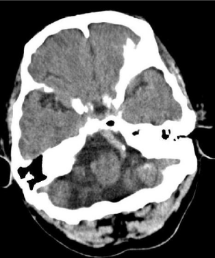
Figure 1: