
Case Report
Austin J Radiol. 2021; 8(4): 1135.
Prenatal Diagnosis of Ductus Arteriosus Aneurysms and Neonatal Outcome: Two Case Reports and Literature Review
Lianza AC*, Morhy SS, Tavares GMP, Gallafrio CC, Sawamura KSS and Fischer CH
Hospital Israelita Albert Einstein, São Paulo, Brazil
*Corresponding author: Alessandro Cavalcanti Lianza, Hospital Israelita Albert Einstein, São Paulo, Brazil
Received: April 14, 2021; Accepted: May 06, 2021; Published: May 13, 2021
Background
Ductus arteriosus aneurysm is a rare condition characterized by a saccular or tubular dilatation of the ductus with potentially fatal outcome due to complications such as rupture, thromboembolism, and infection, compression of adjacent structures and coarctation of the aorta [1]. Association with connective tissue disorders such as Marfan and Ehlers-Danlos syndromes are also described [2,3]. Therefore, we aimed to describe two recent cases of aneurysm of ductus arteriosus assessed by fetal echocardiography and their neonatal outcomes.
Case Presentation
Case 1
A 42-year-old G2P1 woman was referred to fetal echocardiography (fetal 2DE) at 37 weeks of pregnancy (w) due to right cardiac chambers enlargement detected by obstetric ultrasound. The patient referred regular prenatal follow up without any abnormalities or using of medicines besides vitamins. The 2DE fetal demonstrated right cardiac chambers enlargement (primarily related to gestational age) and an aneurysm of the ductus arteriosus with normal ductal flow velocities (Figure 1, Video clip 1). She was admitted in labor at 38 5/7 w. The neonate (female, weight of birth: 2.890g, APGAR 9/10) was delivered without complication. She was transferred to the Neonatal Intensive Care Unit (NICU) for tachypnea. Transthoracic echocardiogram was performed 2 hours after deliver and a foramen ovalis, aneurysm of ductus arteriosus with right to left shunt, thus indicating elevated pulmonary pressures (Figure 2) and a normal-sized Aortic arch was seen. Spontaneous ductus arteriosus occurred 3 days later, without evidence of thrombus or coarctation of the Aorta.
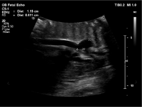
    Figure 1: Bidimensional fetal echocardiography in a sagittal plane of the
ductal arch, demonstrating aneurysm of the ductus arteriosus (arrow).
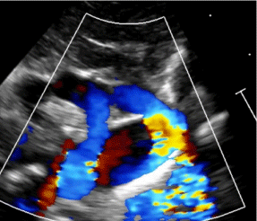
    Figure 2: Doppler evaluation demonstrating aneurysmatic ductus arteriosus
(arrow) and blood flow from the pulmonary artery to the descending aorta
after delivery of the fetus in Figure 1.
Case 2
A 36-year-old G3P2 woman was referred to a routine fetal 2DE at 32w. Prenatal follow up was normal. No medication besides vitamins was being used. The screening demonstrated normal cardiac anatomy and an aneurysm of the ductus arteriosus without ductal flow disturbances (Figure 3a and 3b). She was admitted in labor at 38w and a C-section was indicated due to labor dystocia. The newborn (female, 3.010g, Apgar 9/10) was delivered without complications. Echo was performed in the nursery in the first hour of life. A foramen ovalis, aneurysm of ductus arteriosus with bidirectional shunt (Video clip 2) and an Aortic isthmus diameter below normal range was detected (3.5mm; Z score: -2.39). The neonate was transferred to the NICU for monitorization. Another echo was performed 3 days later, with spontaneous ductal closure, normalization of the Aortic isthmus diameters and no signs of thrombus or coarctation of the aorta (Figure 4).
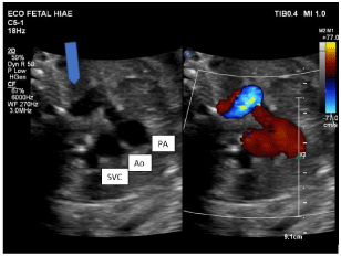
    Figure 3a: Bidimensional fetal echocardiography in a three-vessel view,
demonstrating aneurysm of the ductus arteriosus (arrow). Ao: Aorta; PA:
Pulmonary Artery; SCV: Superior Cava Vein.
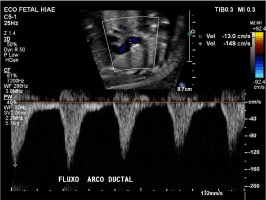
    Figure 3b: Normal fetal ductus arteriosus flow by Doppler analysis in a fetus
with aneurysms of ductus arteriosus.
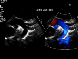
    Figure 4: Echocardiographic image in a suprasternal plane, showing normalsized
Aortic arch after aneurismatic ductus arteriosus closure.
Discussion
We report 2 case reports of ductus arteriosus aneurysms with good clinical outcomes. Of note, only one of the patients needed oxygen supplementation due to tachypnea not related to the cardiac issues, though, symptoms are found in about 11% of the patients in literature [1].
The etiology of these aneurysms in fetal life is unknown, although some theories are found in literature, including (a) congenital weakening of the arterial wall once some authors described necrosis and mucoid degeneration of the media in the arterial wall; (b) presence of increased ductal flow during fetal life secondary to high blood flow as found in arteriovenous shunt and angioma of the liver; in this situation, right chambers enlargement is usually associated [4] and (c) a post stenotic consequence of turbulent flow in fetal ductal constriction [5].
Anatomically, there are three main types of aneurysms od ductus arteriosus: saccular, fusiform and dissecting in which the two formers are considered true aneurysms. Nevertheless, it is unclear which cases should the aneurysm of ductus arteriosus be surgically treated, with the exception of the following conditions: 1) Patent or aneurysmal ductus arteriosus beyond neonatal period; 2) Patients with connective tissue disorder; 3) Thrombus extension into adjacent vessels; 4) Evidence of thromboembolism and 5) Functional compression of adjacent structures [1]. Therefore, in most of cases, spontaneous closure occurs as seen in our cases and no surgical treatment is needed.
Differential diagnosis may include aortic arch abnormalities in fetus; vascular rings in newborns and mediastinal tumors in infants and children. In adults, one may also consider mediastinum tumors, bronchogenic carcinoma and aneurysms of the descending aorta.
Although echocardiography is the most used tool to evaluate these aneurysms and their possible complications, one must consider the operator’s experience and the quality of the images acquired as possible limitations. Imaging the aortic arch by echocardiography, in the presence of aneurysms of ductus arteriosus is challenging, once there may be image overlapping and we may find coarctation of the aorta after the closure of the aneurysm due to potential retraction of the ductal tissue, so we recommend review the aortic arch after the closure of the aneurysm.
Conclusion
Echocardiography is the method of choice for diagnosing and monitoring fetuses and neonates with aneurysm of ductus arteriosus. Although most of cases present spontaneous closure as seen in our institution, we should monitor and be aware of its potential and serious complications.
References
- Dyamenahalli U, Smallhorn JF, Geva T, et al. Isolated ductus arteriosus aneurysm in the fetus and infant: a multi-institutional experience. J Am Coll Cardiol. 2000; 36: 262-269.
- Amato JJ, Cardarelli MG, Bierman FZ. Aneurysm of the arterial duct-a case report and review of the literature. Cardiol Young. 1994; 4: 87-89.
- Crisfield RJ. Spontaneous aneurysm of the ductus arteriosus in a patient with Marfan’s Syndrome. J Thorac Cardiovasc Surg. 1971; 62: 243-247.
- Lund JT, Jensen MB, Hjelms E. Aneurysm of the ductus arteriosus. Eur J Cardiothorac Surg. 1991; 5: 566-570.
- Acherman RJ, Siassi B, Wells W, et al. Aneurysm of the ductus arteriosus: a congenital lesion. Am J Perinatol. 1998; 15: 653-659.