Clinical Image
Austin J Radiol. 2021; 8(4): 1136.
Foramen Magnum Stenosis in Achondroplasia
Choayb S*, Adil H and El Haddad S
Children’s Hospital, Radiology Department, Mohamed V University, Rabat, Morocco
*Corresponding author: Safaa Choayb, Children’s Hospital, Radiology Department, Mohamed V University, Rabat, Morocco
Received: April 24, 2021; Accepted: May 25, 2021; Published: June 01, 2021
Keywords
Achandroplasia; Foramen magnum stenosis; Squamous occipital bone
Clinical Image
Achondroplasia is the most common hereditary skeletal dysplasia and is characterized by disproportionately short stature with rhizomelic short extremities [1]. The skull features include a narrowed foramen magnum, short skull base, and clivus [2].
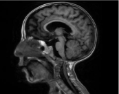
Foramen magnum stenosis is a characteristic funding, secondary to an abnormal placement and premature fusion of the posterior synchondroses [1]. The second factor responsible for stenosis is a defect in endochondral ossification in the basiocciput that may result in an extension of the squamous occipital bone [2]. It can cause hydrocephalus and prominent emissary and meningeal veins (Figure 1).

    Figure 1: Sagittal T1WI revealing a narrowed stenosis of the foramen
magnum and compression of the cervicomedullary junction.
The most severe complication is the compression of the cervicomedullary junction, associated with severe morbidity and sudden death in younger children [1].
Author Contributions
All authors contributed equally to this work.
References
- Sarioglu FC, Sarioglu O and Guleryuz1 H. Neuroimaging and calvarial findings in achondroplasia. Pediatr Radiol. 2020; 50: 1669-1679.
- Caratella S, Tarazi M, Tomalieh FT, Spink G, Bukhari SUA, Ahmad IH, et al. Cranio-cervical junction malformation causing cord compression in infant with achondroplasia: a bigger picture. Br. J. Neurosurg. 2019: 1-3.