Clinical Image
Austin J Radiol. 2021; 8(5): 1138.
Heterotopic Ossifications of the Forearm: A Cause of Post-Traumatic Loss of Pronation-Supination
Hajar A*, Khadija L, Jamal EF and Issam E-N
Department of Radiology, Mohammed V Military Teaching Hospital, Faculty of Medicine and Pharmacy, Mohammed V University, Rabat, Morocco
*Corresponding author: ADIL Hajar, Department of Radiology, Mohammed V Military Teaching Hospital, Faculty of Medicine and Pharmacy, Mohammed V University, Rabat, Morocco
Received: May 14, 2021; Accepted: June 08, 2021; Published: June 15, 2021
Abbreviations
HO: Heterotopic Ossifications; CT: Computed Tomography; R: Radius; U: Ulnar
Keywords
Heterotopic ossifications; Radioulnar; Forearm; Pronationsupination
Clinical Image
HO is defined by the development of ectopic mature bone within nonosseous tissues. It is a well-described phenomenon that complicates forearm fractures, especially when there is an open fracture, a significant soft tissue injury, and associated neural axis or thermal injury. HO mainly forms near metal hardware and may lead to the formation of radio-ulnar synostosis.
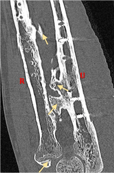
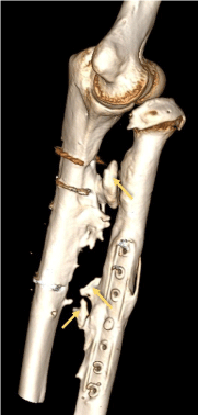
CT is superior to plain radiographs, as it identifies the ectopic bone earlier, defines its exact localization, and helps planning the surgical intervention. Radiologic features are variable; in the early stage, CT shows a low-attenuation mass with indistinct surroundings. As the ossification process progresses, zones of mineralization are visible before leading to the formation of mature cortical bone at the periphery (Figure 1 and 2: arrows). Hastings classification describes 5 classes according to how HO affects the forearm range of motion.

    Figure 1: Coronal oblique CT scan image showing mature bone structures in
the interosseous space and the radio humeral joint.
