Clinical Image
Austin J Radiol. 2021; 8(5): 1139.
Hemifacial Microsomia in an Adult
Hajar A¹*, Laila J¹ and Laamrani FZ²
¹Department of Emergency, Ibn Sina University Hospital, Faculty of Medicine, Mohammed V University, Rabat, Morocco
²Department of Radiology, Ibn Sina University Hospital, Faculty of Medicine, Mohammed V University, Rabat, Morocco
*Corresponding author: Adil Hajar, Department of Emergency, Ibn Sina University Hospital, Faculty of Medicine, Mohammed V University, Rabat, Morocco
Received: May 15, 2021; Accepted: June 10, 2021; Published: June 17, 2021
Keywords
Hemifacial Microsomia; Microtia; Middle ear malformation
Clinical Image
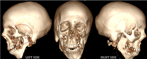
Hemifacial Microsomia is characterized by unilateral underdevelopment of the craniofacial skeleton, external ear, and facial soft tissues. It is the second most common facial birth defect after clefts. HFM is mainly unilateral with a predilection for the right side. In 55% of cases, HFM is associated with extracranial anomalies (Figure 1 and 2).

Figure 1: Overall survival, autologous stem cell transplant (ASCT) versus no ASCT (p=0.12).

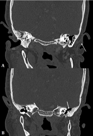
    Figure 2: Coronal CT scan images showing microtia with absence of middle
ear ossicles and external auditory canal atresia (arrows).
CT is the key imaging technique to assess HFM and help evaluate the extent of associated deformities. CT features variably include:
• Shortened mandibular ramus with mandibular condyle hypoplasia associated with chin deviation towards the affected side. The temporomandibular joint deformity is variable.
• Reduced height of the maxilla, zygomatic hypoplasia.
• Microtia or anotia with malformation of the middle ear, lack of pneumatization of the mastoid air cells.
• Orbital malformation, facial musculature hypoplasia may be associated.