Clinical Image
Austin J Radiol. 2021; 8(5): 1140.
Mesenteric Lipoma as an Uncommon Cause of Abdominal Discomfort
Hajar A*, Safaa C, Jamal EF and Mohamed A
Department of Radiology, Mohammed V Military Teaching Hospital, Faculty of Medicine and Pharmacy, Mohammed V University, Rabat, Morocco
*Corresponding author: Adil Hajar, Department of Radiology, Mohammed V Military Teaching Hospital, Faculty of Medicine and Pharmacy, Mohammed V University, Rabat, Morocco
Received: May 15, 2021; Accepted: June 10, 2021; Published: June 17, 2021
Keywords
Lipoma; Mesenteric Lipoma; Abdominal discomfort
Clinical Image
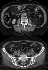
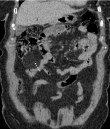
Lipoma is the most frequent benign mesenchymal tumor that resembles normal white fat. Gastrointestinal tract lipomas are rare. The small bowel is the second predilection site of lipomas following the colon. Mesenteric lipomas mainly occur in adults without gender predilection. They are usually asymptomatic and discovered incidentally. However; these tumors may present with intussusception and intestinal bleeding. CT is the key imaging modality to diagnose mesenteric lipoma. They typically present as well-circumscribed, non-enhancing masses with homogeneous fatty attenuation, which are often contained and separate from free mesenteric fat (Figure 1 and 2: white arrows). On MRI, mesenteric lipomas demonstrate homogeneous signal intensity identical to that of fat. Thin fibrous septa of low signal intensity on T1- and T2-weighted images may be present.

    Figure 1: Axial abdominal CT images showing well-circumscribed, nonenhancing
masses with homogeneous fatty attenuation corresponding to
mesenteric lipomas.

    Figure 2: Coronal abdominal CT images showing well-circumscribed, nonenhancing
masses with homogeneous fatty attenuation corresponding to
mesenteric lipomas.