
Clinical Image
Austin J Radiol. 2021; 8(6): 1144.
Retro-Odontoid Pseudotumor Associated with Rheumatoid Arthritis
Hajar A*, Laila J and Laamrani FZ
Department of Emergency, Ibn Sina University Hospital, Faculty of Medicine, Mohammed V University, Rabat, Morocco
*Corresponding author: Adil Hajar, Department of Emergency, Ibn Sina University Hospital, Faculty of Medicine, Mohammed V University, Rabat, Morocco
Received: May 15, 2021; Accepted: June 21, 2021; Published: June 28, 2021
Keywords
Retro-odontoid pseudotumor; Rheumatoid arthritis; Medullary compression
Clinical Image
Retro-Odontoid Pseudotumor (ROP) commonly labeled as “pannus” is a non-neoplastic soft tissue mass adjacent to the odontoid process of C2 that is frequently associated with rheumatoid arthritis. It is due to inflammation and thickening of the synovium surrounding the dens and may lead to ligament laxity, and bone erosion resulting in C1-C2 joint laxity and subluxation with possible spinal cord compression.
MRI is the imaging modality of choice to assess ROP and evaluate its extent. Three patterns of ROP are described following histological features; distinguish inflammatory hyper vascular pannus (hyperintense on T2-weighted images and enhancing) (Figure 1 and 2), combined pannus (intermediate signal intensity on T2- weighted images and intermediately enhancing), and fibrous pannus (low signal intensity on all sequences and non-enhancing).
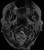
    Figure 1: Axial T2-W cervical spine MRI image demonstrating a hyperintense
retro-odontoid pseudotumor (arrow) compressing the spinal cord (Asterix)
without signs of myelopathy.
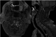
    Figure 2: Axial and sagittal post contrast T1-W FAT SAT cervical spine MRI
images demonstrating peripheral enhancement of the rheumatoid pannus
(arrows).
The images bellow were obtained on a 60 years-old patient with history of RA, who presented with progressive extremities weakness and numbness.