Clinical Image
Austin J Radiol. 2021; 8(6): 1144.
Pulmonary Embolism Saddle
Behyamet O*, Daoud MA, Boris AA, Rachida L and Youssef O
Department of Radiology, National Institute of Oncology, Ibn Sina, Rabat, Morocco
*Corresponding author: Onka Behyamet, Department of Radiology, Mohamed V University, National Institute of Oncology, Ibn Sina Hospital, Rabat, Morocco
Received: May 28, 2021; Accepted: June 24, 2021; Published: July 01, 2021
Keywords
Embolism pulmonary; Saddle; Thoracic angio-CT
Clinical Image
Pulmonary embolism remains a fatal and frequent complication of thromboembolic disease despite the development of preventive methods. Cancer patients are at higher risk of thromboembolism than those in the general population [1]. The thoracic CT angiography is the standard examination; it makes the diagnosis with certainty by showing the endoluminal thrombus. Saddle pulmonary embolism is a radiological term; it is defined by the presence of a thrombus overlapping the bifurcation of the main pulmonary artery extending to both right and left. It represents 2 to 5% of pulmonary embolisms [2].
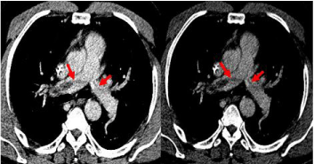
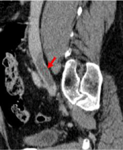
We present the image of a hemodynamically stable 69-year-old patient followed for adenocarcinoma of the prostate who was referred in our training to a thoraco-abdomino-pelvic scanner for assessment and evaluation of his pathology. The chest CT revealed a hypo dense endoluminal thrombus of the pulmonary artery trunk extended to its right and left dividing branches (Figure 1). Abdominal sections showed an endoluminal thrombus of the right common iliac vein extending to the inferior vena cava (Figure 2).

    Figure 1: Chest CT scan with injection of the contrast product in the portal
phase in axial section showing an intraluminal thrombus of the bifurcation
of the pulmonary artery with extension to its left and right dividing branches
producing a saddle appearance (Red Arrow).

    Figure 2: Abdominal CT scan with injection of the contrast product in the
portal phase in sagittal reconstruction showing an intraluminal thrombus of
the right common iliac vein extended to the inferior vena cava (Red Arrow).
The appearance in the saddle is not a sign of seriousness and does not require aggressive therapy. Most patients are hemodynamically stable and respond to standard management of pulmonary embolism with anticoagulants [3].
References
- Prentice A, Ruiz I, Weeda ER. Saddle pulmonary embolism and in-hospital mortality in patients with cancer. International Journal of Clinical Oncology. 2019; 24: 727-730.
- Pathak R, Giri S, Karmacharya P, Aryal MR, Donato AA. Comparison between saddle versus non-saddle pulmonary embolism: insights from nationwide inpatient sample. International journal of cardiology. 2015; 180: 58-59.
- Alkinj B, Pannu BS, Apala DR, Kotecha A, Kashyap R, Iyer VN. Saddle vs Nonsaddle Pulmonary Embolism: Clinical Presentation, Hemodynamics, Management, and Outcomes. Mayo Clinic proceedings. 2017; 92: 1511- 1518.