Clinical Image
Austin J Radiol. 2021; 8(6): 1147.
Focal Hepatic Hot Spot Sign
Mohamed DA*, Retal H, Onka B, Latib R and Omor Y
National Institute of Oncologic, University Hospital Center Ibn Sina, Rabat, Morocco
*Corresponding author: Daoud Ali Mohamed, Radiology Department, University Hospital Center Ibn Sina, Rabat, Morocco
Received: June 04, 2021; Accepted: July 06, 2021; Published: July 13, 2021
Abstract
The focal hepatic hot spot sign appears as an area of increased radiopharmaceutical uptake of the quadrate lobe of the liver in the arteial an veinous phase. This sign seen on CT is due to obstruction of the superior vena cava and portosystemic venous shunt between the superior vena cava and the left portal vein via the thoracic and internal para-umbilical veins.
Keywords: Hepatic hot spot; CT scan; Hemangioma
Clinical Image
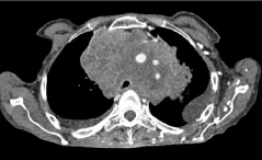
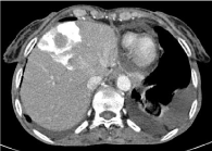
A 55-year-old woman followed for right plantar melanoma with secondary localization (cerebral, thoracic and abdominal) treated with chemotherapy. Three months later, the CT scan showed an increase of the mediastinal mass compressing the mediastinal vascular structures, in particular the superior vena cava (Figure 1). CT of the abdomen revealed an intense wedge-shaped homogeneous enhancement area in the quadrate lobe of the liver (Figure 2).

    Figure 1: Axial contrast enhanced CT of the chest showing a mediastinal
mass (metastasis of a melanoma) compressing the vascular structures with
obstruction of the superior vena cava.

    Figure 2: Axial contrast enhanced CT of the abdomen showing an intense
wedge-shaped enhancement area in the quadrate lobe of the liver in late
arterial phase : sign of the focal hepatic hot spot sign.
Discussion
The focal hepatic hot spot sign was first described in 1983 by Ishikawa. The sign can be observed on technetium 99m (99mTc) Sulphur colloid scans or on contrast, material enhanced CT scans [1]. This abnormal enhancement is due to portosystemic venous shunting between the SVC and portal vein. The hot spot is created by areas of focally increased blood flow that result from this shunting [2].
This sign has been reported in Budd-Chiari syndrome, the causes of SVC syndrome (neoplasms of the thorax as lung carcinoma and lymphoma, Vasculo-Behcet’s disease, fibrosing mediastinitis, and luetic aneurysm) [3].
There are other conditions which can result in increased uptake such as hemangioma, hepatocellular carcinoma, focal nodular hyperplasia, which if they occur in regions adjacent to the falciform ligament in segment IV can mimic the focal hepatic hot spot sign.
References
- Dickson AM. The focal hepatic hot spot sign. Radiology. 2005; 237: 647-648.
- Hoang VT, Vo NQ, Trinh CT, et al. The focal hepatic hot spot sign with lung cancer in computed tomography. Respirol Case Rep. 2020; 8: e00671.
- Phoophiboon V, Tantiprawan J, Vanakiatkul H and Wongkarnjana A. Systemic to pulmonary venous shunt and the focal hepatic hot spot sign from SVC obstruction in Behcet’s disease. BMJ Case Rep. 2020; 13: e234017.