Case Report
Austin J Radiol. 2021; 8(7): 1152.
Man in the Barrel Syndrome Following TEVAR
Biao Zhi¹, Xiangke Niu¹, Yong Chen²*
1Department of Radiology, Affiliated Hospital of Chengdu University, Chengdu, Sichuan, China
2Department of Intervention, Affiliated Southern Hospital of Southern Medical University, Guangzhou, Guangdong, China
*Corresponding author: Yong Chen, Department of Intervention, Affiliated Southern Hospital of Southern Medical University, Guangzhou, Guangdong, China
Received: July 10, 2021; Accepted: August 04, 2021; Published: August 11, 2021
Abstract
Man-in-the-Barrel Syndrome (MIBS) is a neurological disorder characterized by the paralysis of both upper limbs without paralysis of both lower limbs or paralysis of the pathological reflex and is very rare in clinical practice. The pathogenesis of MIBS varies and includes disorders of the brain, brainstem, spinal cord or peripheral nerves. Most cases are due to intracranial lesions, and MIBS caused by cervical spinal cord ischemia is particularly rare. This study reports a case of MIBS caused by cervical spinal cord ischemia one day after Thoracic Endovascular Aortic Repair (TEVAR).
Keywords: Man-in-the-barrel syndrome (MIBS); Aortic dissection; TEVAR; Cervical spinal cord ischemia; Fenestration operation
Case Presentation
This retrospective study was compliant with the Health Insurance Portability and accountability act and approved by our institutional review board, which waived the requirement for written informed consent.
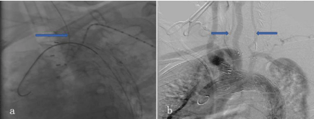
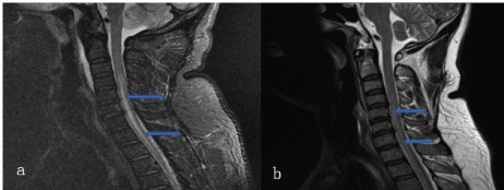
A 47-year-old male patient was admitted to the hospital on March 5, 2019, for sudden severe chest and back pain with left lower extremity pain. CT examination revealed aortic dissection. The dissection involved the opening of the left common carotid artery and occlusion of the left lower extremity artery. On the night of admission, thoracic aortic stenting, after the patient and his wife signed the informed consent, left common carotid artery stent implantation by fenestration and left iliac artery stenting were performed under general anesthesia. Intraoperative angiography showed that the descending aorta had a tera and a true and false lumen were observed; both vertebral arteries converged to the basilar artery, and the blood flow was unobstructed (in a balanced pattern) (Figure1b). The main stent (VAMF36C200TE, Medtronic Company) was implanted to cover the opening of the left subclavian artery and left common carotid artery, and the branch stent (FLUENCY, 8mm×40mm, BARD Company) was implanted into the left common carotid artery through a fenestration operation (Figure 1a). The left iliac artery was implanted with an 8mm×100mm covered stent (VIABAHN, Gore Company) and an 8mm×100mm bare stent (E-LUMINEXX, BARD Company). Postoperative ascending aortography reexamination showed that the stent-covered thoracic aorta was securely attached, the blood flow in the left common carotid artery and brachiocephalic trunk artery was unobstructed, and the left subclavian artery was visible. The arterial blood flow in the left lower extremity was unobstructed. The operation was completed at 1:00 on March 6, 2019, and the procedure, resuscitation and extubation were successful. Decreased muscle strength in both upper limbs (Grade II) was noted at 8:00 on March 7, 2019, along with symmetric hypoesthesia. The upper limbs could not be lifted flat and raised to the top of the head; there was decreased grip strength in both hands, with normal movement and sensation in both lower limbs. The right upper limb blood pressure was 143/85 mmHg, and the left upper limb blood pressure was 90/70 mmHg. On March 8, 2019, enhanced cervical spine MRI (3T) showed cervicothoracic spinal cord swelling, an increased gray matter signal, and ischemic changes in addition to the clinical symptoms (Figure 2a). Bilateral upper extremity electromyography showed that F waves of the bilateral median and right ulnar nerve were not elicited. It was suggested that the F waves of the bilateral median and right ulnar nerve were abnormal. According to the clinical manifestations and the results of the auxiliary examinations, the patient was diagnosed with Man-in-the-Barrel Syndrome (MIBS), which was caused by closure of the left subclavian artery. After communicating with the patient and his wife and obtaining consent, a 10mm×40mm stent (FVL10040, BARD Company) was placed in the left subclavian artery by fenestration on the evening of March 8, 2019. Postoperative angiography showed that the blood flow of the left subclavian artery and the left vertebral artery was unobstructed, and no internal leakage was observed (Figure 1b). After the operation, the muscle strength of both upper limbs increased (Grade III), and both upper limbs could be lifted to the top of the head. The sensation in both upper limbs increased significantly, and the grip strength in both hands remained unchanged. The blood pressure in both upper extremities is similar. On March 20, 2019, a cervical spinal cord MRI revealed that the swelling of the cervicothoracic spinal cord and the increase in the gray matter signal were significantly alleviated in consideration of ischemic changes (Figure 2b). At present, the patient continues to undergo rehabilitation.

    Figure 1: a) Left common carotid artery fenestration. The arrow points to the fenestration needle. b) Postoperative angiography, showing that the vertebral artery
was balanced. The arrow points to the bilateral vertebral artery.

    Figure 2: a) Sagittal-T2WI before fenestration of the left subclavian artery. The arrow indicates an abnormal signal in the cervical spinal cord. b) Sagittal-T2WI of
the cervical spinal cord after fenestration of the left subclavian artery. The arrow indicates an abnormal signal in the cervical spinal cord.
Discussion
MIBS was first reported by Sage Equivalent in 1983. MIBS is characterized by the limited active movement of both upper limbs and normal function in both lower limbs. MIBS got its name because the conditions are similar to those that would be experienced by a person whose upper limbs are confined in a barrel. MIBS usually refers to acute bilateral upper limb paralysis caused by cerebral infarction in the anterior and middle cerebral artery watershed caused by hypoperfusion. Bilateral arm paralysis caused by other causes, such as pons, spinal cord and peripheral nerve damage, is also called barrel-man syndrome. Pathologically, MIBS can be caused by the involvement of the upper or lower motor neurons. Upper motor neuron lesions mainly refer to the motor representative areas of the brain that affect the function of the hand and arm, including the pyramidal tracts of the medulla oblongata, while lower motor neuron lesions may affect the anterior horn of the spinal cord, the anterior cervical spinal nerve root, or the brachial plexus. Calle-Lopez Y, et al. [1] reported a case of barrel-man syndrome caused by brachial plexus arteritis. Asmaro K, et al. [2] reported a case of barrel-man syndrome caused by cervical spinal epidural abscess. There are few reports of cervical spinal cord infarction. The common area of ischemic spinal cord injury is the watershed of the back or lumbar spine. The most common cause of MIBS is anterior spinal artery infarction [3]. The anterior spinal artery is composed of two branches of the intracranial segment of the vertebral artery that descend from the ventral side of the spinal cord along the anterior median fissure. The anterior spinal artery branches into the spinal cord to supply blood to the anterior 2/3 of the spinal cord. Infarction in this area may be caused by hypoperfusion of the anterior spinal artery or vertebral-basilar artery. The causes of hypoperfusion of these arteries include vascular stenosis or embolism.
Type III aortic dissection may be partially compensated with the right pyramidal artery by closing the left subclavian artery when the right vertebral artery is dominant or the bilateral vertebral artery is homogeneous and the pyramidal-basilar artery ring is patent. A total of 629 related cases were analyzed by Shu Chang et al. [4], 159 of these cases showed complete occlusion. The results showed that there was no significant difference in the incidence of left subclavian artery paraplegia between occluded and nonoccluded arteries. The occlusion of the left subclavian artery did not increase the risk of long-term cerebral infarction. Even if some could not be fully compensated, the patient had mild symptoms of vertebral artery theft, such as ischemia of the left upper limb, dizziness and fatigue, and the quality of life was not affected. However, although this patient met the aforementioned conditions, MIBS caused by cervical spinal cord ischemia is very rare after left subclavian artery occlusion, which deserves clinical attention. Once symptoms related to cervical spinal cord ischemia occur, the left subclavian artery must be opened immediately. In this case, fenestration was successfully completed by puncturing the TEVAR stent, and the left subclavian artery was opened. No complications such as internal leakage, pneumothorax or hemorrhage were observed. This result suggests that in this case, MIBS may have a result of the decrease in left vertebral artery blood flow caused by the occlusion of the left subclavian artery by endovascular aortic dissection repair and the hypoperfusion of the anterior spinal artery and vertebral-basilar artery.
References
- Calle-Lopez Y, Fernandez-Ramirez AF, et al. Man-in-the-barrel syndrome: atypical manifestation of giant cell arteritis. Rev Neurol. 2018; 66: 373-376.
- Asmaro K, Pabaney AH, et al. Man-in-the-barrel syndrome: Case report of ventral epidural abscess and review of the literature. Surg Neurol Int. 2018; 9: 8.
- Gonzalez-Usigli H, Gandarilla A, et al. Cervical ischaemic neuronopathy and cardioembolism: another cause of man-in-the-barrelsyndrome. Rev Neurol. 2016; 63: 543-546.
- Shu C, WAng SL, et al. Safety of left subclavian artery coverage during thoracic endovascular aortic repair. Chinese Journal of General Surgery. 2014; 23: 1614-1619.