Clinical Image
Austin J Radiol. 2021; 8(8): 1154.
Early Cortical Laminar Necrosis on MRI
Mohamed DA*, Onka B, Choayb S, En-Nafaa I, Fenni JEL and En-Nouali H
Department of Radiology, Mohamed V Military Hospital, Rabat, Morocco
*Corresponding author: Daoud Ali Mohamed, Department of Radiology, University Hospital Center Ibn Sina, Rabat, Morocco
Received: July 19, 2021; Accepted: August 12, 2021; Published: August 19, 2021
Clinical Image
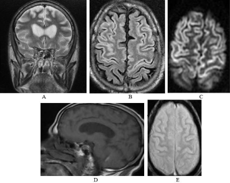
An 80-year-old man with chronic hypertension was admitted to the emergency department with consciousness disorders. The evolution was marked by a rapid worsening of his neurological condition. The patient was intubated and ventilated. The biological check-up revealed a blood glucose level of 0.2g/l. A brain scan was performed which was without abnormality. Two days after the normalization of the blood sugar level, the patient presented a late awakening. A brain MRI was performed which showed bilateral fronto-parietal laminar cortical areas in T2, Flair and diffusion hypersignal, T1 iso signal, and no hyposignal on T2 gradient echo sequence (Figure 1). The diagnostic of Cortical Laminar Necrosis was retained.

    Figure 1: Coronal T2 (A), axial Flair (B), and axial diffusion (C) Showed
hyperintense bilateral fronto-parietal laminar cortical. Sagittal T1 showed (D)
Iso signal and no hyposignal on axial T2 gradient echo sequence (E).
Discussion
Cortical laminar necrosis corresponds to neuronal ischemic remodeling secondary to hypoxia. Grey matter is more vulnerable than white matter; it can be affected in isolation, which defines “selective neuronal necrosis” [1]. The third cortical layer is the most sensitive. If the energy deficit continues, more extensive brain damage occurs, affecting all parenchymal structures of the brain [2]. Histologically, laminar cortical necrosis corresponds to a neuronal loss preferentially affecting layers III and V. Depending on the intensity of the damage, the necrosis of the cortex is more or less extensive and is associated with the presence of macrophages, gliosis and vascular neogenesis [3]. The aetiologies are variable and are represented by intoxication, hypoglycaemia, ischaemia, prolonged arterial hypotension, status epilepticus, cardiac and respiratory arrest cardiac and respiratory arrest, renal or hepatic dysfunction, immunological causes immunological causes, as in antiphospholipid syndrome or lupus, and infectious involvement in encephalitis [1,2,4]. The clinical picture is dominated by disorders of consciousness, sometimes a motor or sensory deficit [1]. MRI is the examination of choice for the diagnosis of cortical laminar necrosis and for the accuracy of lesion location and extent [2,3]. Sawada et al reported the first MRI description of NCL in 1990 and then several authors described this lesion with more precision. Lesions appear hypersignal on diffusion, T2-weighted and T2 Flair sequences within the first 24 hours and for the first three weeks [5]. The T1 sequence shows hyposignal or iso-signal brain oedema during the first two weeks, followed by spontaneous cortical and basal ganglia hypersignal after two weeks. T2 gradient echo sequences do not show hyposignal of the cortical lesions, thus ruling out a haemorrhagic origin. The prognosis is poor, with high morbidity and mortality. There is no specific curative treatment, which depends mainly on the aetiology [2].
References
- Siskas N, Lefkopoulos A, Ioannidis I, Charitandi A, Dimitriadis AS. Cortical laminar necrosis in brain infarcts: serial MRI. Neuroradiology. 2003; 45: 283- 288.
- Donaire A, Carreno M, Gómez B, Fossas P, Bargalló N, Agudo R, et al. Cortical laminar necrosis related to prolonged focal status epilepticus. J Neurol Neurosurg Psychiatry. 2006; 77: 104-106.
- Komiyama M, Nakajima H, Nishikawa M, Yasui T. Serial MR observation of cortical laminar necrosis caused by brain infarction. Neuroradiology. 1998; 40: 771-777.
- Rajasekharan C, Jithesh B, Renjith SW. Cortical laminar necrosis due to hypoglycaemic encephalopathy: images in medicine. BMJ Case Rep. 2013.
- Yoneda Y, Yamamoto S. Cerebral cortical laminar necrosis on diffusionweighted MRI in hypoglycaemic encephalopathy. Diabet Med. 2005; 22: 1098-1100.