
Case Report
Austin J Radiol. 2021; 8(9): 1162.
Atypical 99mTc-HMDP Distribution in a Patient with Poorly Differentiated Laryngeal Carcinoma: A Case of Paraneoplastic Hypercalcemia
Altini C1, Rizzo A2* and Bruno I3
1Nuclear Medicine Unit/Imaging Department, IRCCS Bambino Gesù Children’s Hospital, Piazza Sant’Onofrio 4, 00165, Rome, Italy
2Candiolo Cancer Institute, FPO - IRCCS, Candiolo, TO, Italy
3Unità Operativa Complessa di Medicina Nucleare, Fondazione Policlinico Universitario A. Gemelli IRCCS, Roma, Italy
*Corresponding author: Alessio Rizzo, Candiolo Cancer Institute, FPO - IRCCS, Candiolo, TO, Italy
Received: July 29, 2021; Accepted: September 23, 2021; Published: September 30, 2021
Abstract
A 55 years-old man underwent Whole-Body Computed Tomography (CT) in suspicion of laryngeal carcinoma. The CT highlighted a voluminous neck mass with epicenter in the epiglottic region and latero-cervical lymph-nodal involvement. To assess the presence of bone metastasis and to complete the staging, the patient underwent a whole-body bone scintigraphy with 99mTc- HMDP. At the time of the exam, a significant hypercalcemia was detected. The planar scan showed massive 99mTc-HMDP uptake in liver, lungs, heart and pancreas, compatible with the presence of microcalcifications. The laryngeal biopsy evidenced a squamous poorly differentiated laryngeal carcinoma, justifying the functional findings previously reported.
Keywords: 99mTc-HMDP; Computed tomography; Laryngeal carcinoma
Case Presentation
A 55 years old man, long-time smoker, was hospitalized for a swelling in the right side of the neck. The analysis performed during the hospitalization highlighted a calcemia of 18.3mg/dL, an alkaline phosphatase of 224UI/L, a PTH of 8.7pg/mL, a 25-OH Vitamin D of 15.5ng/mL and a creatinine of 2.39mg/dL; ultrasound showed a mass in the right side of larynx and some suspicious lymph-nodes.
Subsequently he underwent a Whole-Body Computed Tomography (CT) that highlighted a voluminous mass in the right side of the neck, in the epiglottic-suprahyoid region with evidence windpipe deviation and right latero-cervical lymph-nodal involvement; the exam did not show any other peculiar finding.
The following neck magnetic resonance confirmed CT findings specifying the epicenter of the mass in the right supra-glottic region.
The carcinoma of the larynx is the most frequent upper aerodigestive tract tumour, its incidence in higher in 50-60 years old men and in more than 95% show squamous [1] histopathologic pattern. In order to assess the presence of bone involvement and to complete the staging, the patient underwent a bone scintigraphy in our department.
Whole-body bone scan was performed two hours after intravenous injection of 99mTc-HMDP (administered activity: 546MBq) as recommended by EANM guidelines for bone scintigraphy [2]. Scintigraphy images (A: Anterior view; B: Posterior view) (Figure 1) showed a massive uptake of the tracer in the liver (black arrow) and, to a lesser extent but still significant, in lungs, heart and pancreas; these findings, considering the absence of any other clinical reason [3] (e.g. ATTR cardiac amyloidosis, hyperparathyroidism), are compatible with the presence of microcalcifications in the soft tissues. Bone scan images did not display any sign of abnormal distribution of 99mTc-HMDP due to the presence of bone metastases. SPECT-CT was not performed due to patient’s poor clinical conditions (overall the mental confusional state). Laryngeal biopsy evidenced a squamous poorly differentiated laryngeal carcinoma.
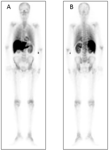
Figure 1:
Thus, the patient has been classified as cT3N2bM0 (Stage IVa; UICC/AJCC 8th edition).
Patients with this stage of disease have different chances of treatment: total laryngectomy +/- lynphadenectomy followed by adjuvant treatment (radiotherapy or chemo-radiotherapy). If the clinical status of the patient does not allow a surgical approach or the tumour is unresectable, a neoadjuvant chemo-radiotherapy treatment should be considered, keeping the surgery as a second chance in case of failure.
Our patient underwent a total laryngectomy and bilateral laterocervical lymph-node dissection.
The definitive histological examination confirmed the diagnosis of squamous poorly differentiated laryngeal carcinoma with nodal metastasis.
Hypercalcemia can be a manifestation of advanced squamous cell carcinoma of the head and neck cancer. Moreover, hypercalcemia without bone metastases is presumably due to the production of ectopic parathormone-related protein (PTH-rP) from tumour cells4, justifying our scintigraphic findings. The association between hypercalcaemia and head and neck squamous cancer is uncommon and the incidence of hypercalcaemia in this kind of disease is suggested to be higher at the advanced stages [5-8].
References
- AIOM Guidelines.
- T Van den Wyngaert, K Strobel, WU Kampen, T Kuwert, W van der Bruggen, HK Mohan, et al, On behalf of the EANM Bone & Joint Committee and the Oncology Committee. The EANM practice guidelines for bone scintigraphy. Eur J Nucl Med Mol Imaging. 2016; 43: 1723-1738.
- Wale DJ, Wong KK, Savas H, Kandathil A, Piert M, Brown RK. Extraosseous Findings on Bone Scintigraphy Using Fusion SPECT/CT and Correlative Imaging. AJR Am J Roentgenol. 2015; 205: 160-172.
- Sellers RS, Schuller DE, Sharma PK, et al. Head and neck squamous cell carcinoma; Measurement of plasma parathyroid hormone-related protein and serum and urine calcium concentrations. Otolaryngol Head Neck Surg. 2000; 123: 558-562.
- Patrick J. Bradleya and David Hoskinb. Hypercalcaemia in head and neck squamous cell carcinoma. Curr Opin Otolaryngol Head Neck Surg. 2006; 14: 51-54.
- Sridhar KS, Hussein AM. Hypercalcemia in head and neck squamous-cell carcinoma. Am J Clin Oncol. 1990; 13: 388-593.
- Victor M Gastanaga, Lee S Schwartzberg, Rajul K Jain, Melissa Pirolli, David Quach, Jane M Quigley, et al. Prevalence of hypercalcemia among cancer patients in the United States. Cancer Med. 2016; 5: 2091-2100.
- Iwase M, Takemi T, Manabe M, Nagumo M. Hypercalcemic complication in patients with oral squamous cell carcinoma. Int J Oral Maxillofac Surg. 2003; 32: 174-180.