Case Report
Austin J Radiol. 2021; 8(10): 1167.
Kidney Stones Vanished into Thin Air: Distinguishing Emphysematous Pyelitis from Renal Calculi on Ultrasound
Rajendram R1,2*, Syed Y3, Bakhsh UR3 and Hussain A4
1Department of Medicine, King Abdulaziz Medical City, King Abdullah International Medical Research Center, Ministry of National Guard - Health Affairs, Riyadh, Saudi Arabia
2College of Medicine, King Abdulaziz University for Health Sciences, Riyadh, Saudi Arabia
3Department of Intensive Care Medicine, King Abdulaziz Medical City, King Abdullah International Medical Research Center, Ministry of National Guard - Health Affairs, Riyadh, Saudi Arabia
4Department of Cardiovascular Sciences, King Abdulaziz Medical City, King Abdullah International Medical Research Center, Ministry of National Guard - Health Affairs, Riyadh, Saudi Arabia
*Corresponding author: Rajkumar Rajendram, Department of Medicine, King Abdulaziz Medical City, Ministry of National Guard - Health Affairs, Riyadh, Saudi Arabia
Received: September 23, 2021; Accepted: October 06, 2021; Published: October 13, 2021
Abstract
Emphysematous pyelitis (gas within the excretory system) is a rare infection with a mortality of 20%. Although, it may resolve with antibiotics alone, the presence of gas within the kidney must be detected and clearly distinguished from uncomplicated pyelonephritis or calculi. Acoustic shadowing on ultrasound (i.e. the drop in echo strength under a highly reflective or attenuating structure) can be useful in this situation. This can distinguish calcification (clean shadowing) or bone (partial shadowing) and gas (dirty shadowing). To highlight the importance of the correct interpretation of this imaging artifact we describe the course of a 64 years old woman who presented with fever, left flank pain and dysuria. The renal tract ultrasound was initially reported to show multiple renal calculi. However, on re-review it was noted that the appearance of the acoustic shadowing was more consistent with air. The diagnosis of emphysematous pyelitis was confirmed by computed tomography.
Keywords: Emphysematous pyelitis; Pyelonephritis; Ultrasound; Acoustic shadowing; Renal calculi
Introduction
Albeit rare, emphysematous pyelitis is associated with significant mortality. Yet, it may resolve with antibiotics alone [1]. With a mortality approaching 50%; emphysematous pyelonephritis (gas within the renal parenchyma) requires drainage or nephrectomy [1]. Regardless, the presence of gas within the kidney must be detected and clearly distinguished from uncomplicated pyelonephritis or calculi. As illustrated by the case report below, this can be achieved with ultrasound; a non-invasive test that can be performed at the patient’s bedside.
Case Report
A 64-year-old woman presented with a three days history of intermittent left flank and abdominal pain preceded by a 10 days history of fevers, dysuria and foul-smelling urine. Her past medical history included diabetes mellitus (metformin 500mg three times per day, gliclazide 120mg daily, sitagliptin 100mg daily), hypothyroidism (levothyroxine 25μg daily), hyperlipidaemia (atorvastatin 40mg daily), hypertension (amlodipine 5mg daily, perindopril 2.5mg daily, indapamide 1.5mg daily), right cerebellar stroke (aspirin 81mg daily) and lumbar spinal canal stenosis (carbamazepine 400mg daily).
Besides intermittent high-grade fevers, vital signs were unremarkable. Physical examination revealed only left flank tenderness. Laboratory blood results revealed hyperglycaemia (glucose 23.8mmol/l), lactic acidosis (lactate 5.31mmol/l) and stage 1 acute kidney injury (creatinine 138μmol/l, baseline 97μmol/l; urea 13.5mmol/l).
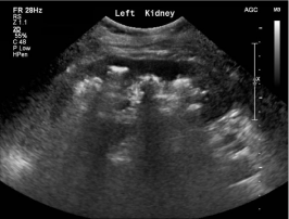
A renal tract ultrasound was reported to show multiple stones of varying sizes in the left kidney with mild hydroureteronephrosis (Figure 1) and echogenic debris within the bladder. However, on re-review of the images, it was recognised that the ‘dirty’ acoustic shadows’ (i.e., echogenic foci with several reverberation artifacts) seen in the left kidney (Figure 1) were more consistent with air. Renal calculi usually produce partial or ‘clean’ acoustic shadows [2]. The diagnosis of emphysematous pyelitis (gas within the pelvicalyceal system without parenchyma involvement) was confirmed by unenhanced Computed Tomography (CT) of the abdomen (Figure 2).

    Figure 1: Ultrasound scan of the left kidney.
Legend to Figure 1. The renal tract ultrasound was initially reported
to show multiple stones of varying sizes in the left kidney with mild
hydroureteronephrosis. However, on re-review, the ‘dirty’ acoustic shadows’
(i.e. echogenic foci with several reverberation artifacts) seen in the left kidney
were more consistent with air. Renal calculi usually produce partial or ‘clean’
acoustic shadows [2].

    Figure 2: Unenhanced computed tomography scan of the kidneys, ureters
and bladder.
Legend to Figure 2. This coronal image from an unenhanced computed
tomography scan of the kidneys, ureters and bladder demonstrated gas
within the left pelvicalyceal system without parenchymal involvement. This
confirmed the diagnosis of emphysematous pyelitis.
Urine and blood cultures grew extended spectrum beta-lactamase producing Escherichia coli. The fevers, dysuria, flank pain, renal function and inflammatory markers improved with intravenous meropenem. Abdomen CT, repeated one week after admission, demonstrated almost complete resolution of the emphysema. The next day the patient insisted upon discharged home, against medical advice. So, oral ciprofloxacin was administered for another 7 days.
Discussion
The mortality associated with emphysematous infections of the urinary tract is significantly greater than that of uncomplicated pyelonephritis [1]. Therefore, the presence of gas within the kidney must be detected and clearly distinguished from uncomplicated pyelonephritis or calculi. In this context, acoustic shadowing on ultrasound can be clinically useful. Acoustic shadowing refers to the drop in echo strength under a highly reflective or attenuating structure. The tissue gain compensation fails to adequately amplify these echoes which appear as a band below the object [2]. This can distinguish calcification (clean shadowing) or bone (partial shadowing) and gas (dirty shadowing) [2].
Clean shadowing (a dark anechoic band) occurs if most of the ultrasound energy is absorbed. So, there are few secondary reflections. Larger calculi and bone usually cause clean acoustic shadowing [2]. Renal calculi are difficult to distinguish from adjacent echogenic tissues [2]. Shadowing is therefore important for their identification [2]. Partial shadowing (a hypoechoic band) occurs distal to tissues that highly attenuate ultrasound (e.g., fat, small stones) [2]. Dirty shadowing (Figure 1) occurs when a highly reflective surface (e.g., gas) causes multiple secondary low-level echo reflections (reverberation artifacts) to appear within the shadows [2].
Conclusion
The correct interpretation of acoustic shadowing on ultrasound can distinguish gas, bone and calcification. This can facilitate the diagnosis of emphysematous pyelonephritis and pyelitis.
Learning points
Careful assessment of acoustic shadowing on ultrasound can distinguish air from stones and bone.
Isolated gas within the excretory system (emphysematous pyelitis) must be differentiated from emphysematous pyelonephritis (gas in the parenchyma of the kidney).
Emphysematous pyelitis may resolve with antibiotics alone but emphysematous pyelonephritis requires drainage or nephrectomy.
Sources of funding
This research did not receive any specific grant from funding agencies in the public, commercial, or not-for-profit sectors.
References