Clinical Image
Austin J Radiol. 2021; 8(11): 1168.
LV Apical Aneurysm with Apical Ventricle Septal Rupture Following Anterior Wall Infarction
Hassan A¹*, Khan F² and Azom K¹
1Department of Medicine, Saint Richard’s Hospital Chichester, UK
2Resident Cardiology, Ayub Teaching Hospital Abbottabad, UK
*Corresponding author: Awab Hassan, Department of Medicine, Saint Richard’s Hospital Chichester, UK
Received: September 13, 2021; Accepted: October 22, 2021; Published: October 29, 2021
Clinical Image
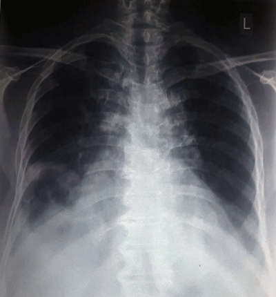
A 49-year-old female patient presented with complaints of progressively increasing shortness of breath and reducing exercise tolerance since the last 4 days. Patient had an anterior wall myocardial infarction 11 days ago and was discharged following an uneventful stay in the coronary care unit. Upon presentation an X-ray was done by the E.R physician, which showed bilateral pleural effusions with focal lateral bulge arising from the left lower heart border, this raised suspicion of ventricular aneurysm.
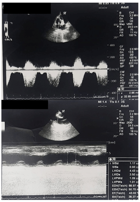
Echocardiography showed anteroseptal and apical akinesia. There was an outpouching in the anterior wall with distal septal rupture; Left to right shunting was observed. ECG showed persistent ST elevation in the precordial leads, which is a classic feature of ventricular aneurysm. After stabilization with diuretics patient was discussed with cardiothoracic surgery team and subsequently referred to them (Figure 1).

Figure 1:
Ventricular aneurysms are a rare but serious complication following ST elevated myocardial infarction. Prevalence rate is 5 percent post infarction, mostly associated with anterior wall infarctions. Most are managed medically; prognosis is good after surgical repair (Figure 2).
