
Clinical Image
Austin J Radiol. 2021; 8(11): 1169.
A Non-Aspergillosis Cause of Air Crescent Sign
El Ouali I*, Elaitari K, Jerguigue H, Latib R and Omor Y
Department of Radiology, Oncology National Institute, Rabat, Morocco
*Corresponding author: El Ouali Ibtissam, Department of Radiology, Oncology National Institute, Lalla Asmaa Avenue, Al Azhar Residency A, Apt 21. Tabriquet, Salé, Morocco
Received: October 12, 2021; Accepted: November 03, 2021; Published: November 10, 2021
Keywords
Air Crescent Sign; Lung Cancer; Chemotherapy
Abbreviations
CT: Computed Tomography
Clinical Image
A 65 old man, with a history of heavy smoking was diagnosed with pulmonary adenocarcinoma. The initial chest CT showed a solid mass in the upper left lobe (Figure 1).
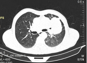
    Figure 1: A chest CT-Scan in axial reconstruction revealing a pulmonary
adenocarcinoma in the left upper lobe prior to chemotherapy.
Post-chemotherapy chest CT revealed a cavitary lesion with an air-crescent sign (Figure 2). The resulting aspect is an expression of the necrotic excavation of the tumor.
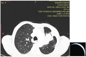
    Figure 2: A chest CT scan in axial reconstruction performed after
chemotherapy, revealing a cavitary lesion with thick wall and irregular outer
border, with an air-crescent sign in the left upper lobe resembling a fungus
ball mass.
The increased granulocyte activity causes the tumor to necrosis, then the liquefied material is evacuated completely or partially
into a bronchus, leaving a residual lumen filled by air, interposed between the devitalized tissue and the surrounding parenchyma, bearing a likeness to a fungus ball mass frequently recognized by its characteristic radiological appearance that we refer to as: the air crescent, meniscus or cap sign [1].
The physician should keep in mind that particular histological types of lung cancer may present an air-crescent sign, spontaneously or after chemotherapy.
References