Case Report
Austin J Radiol. 2021; 8(11): 1170.
Idiopathic EJV Pseudoaneurysm
Saadawy A*
Department of Radiology, West Suffolk Hospital, Addenbrookes Hospitals, UK
*Corresponding author: Ahmed Saadawy, Department of Radiology, West Suffolk Hospital, Addenbrookes Hospitals, UK
Received: October 22, 2021; Accepted: November 11, 2021; Published: November 18, 2021
Abstract
As opposed to arterial pseudoaneurysm, Venous pseudoaneurysms are very rare. Particularly EJV being more rare than IJV. A study published in 2018 found that there is less than 10 well-documented cases of EJV aneurysms accessible.
Keywords: Pseudoaneurysm; Posterior triangle; Headache
Case Presentation
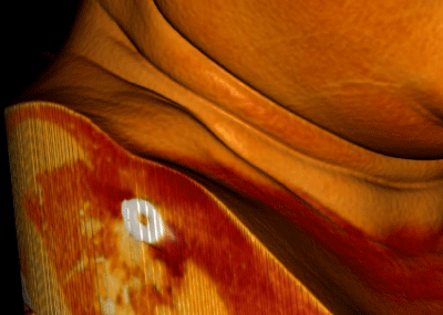
49 y/o lady presenting with a golf-ball sized painless neck swelling in the right lower posterior triangle. The swelling has been increasing in size over a year duration and particularly increases in size when lying down. She was experiencing pressure symptoms and tightness on the ipsilateral side of the face along with a right hemi-cranial headache and retro-orbital pain [1].
Clinical vascular scientist reported the following:
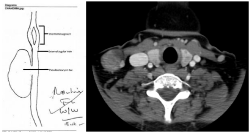
• The lump in the neck corresponds to a pseudoaneurysm of the external jugular vein.
• The defect in the vein wall measures approximately 3mm in dimeter.
• The sac measures 1.3cm (AP) x 2.9cm (Transverse) x 2.4cm (Length)
• Despite very slow movement within the sac, there is no evidence of any thrombosis.
The swelling was excised under local anesthetic in 2015.
Imaging Findings
CT Neck with contrast
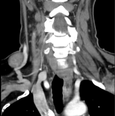
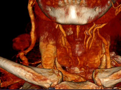
• Showed 2.6 mixed attenuation rounded object in relation to EJV, with a blush of contrast from the External jugular vein into the vascular structure.
• Appearances most in keeping with venous pseudoaneurysm originating from the EJV at the level of the thyroid gland.
Handheld doppler
• No arterial signal (Figure 1-4).

Figure 1:

Figure 2:

Figure 3:

Figure 4:
Discussion
A Pseudoaneurysm is defined according to Mayo clinic as an injury to the vessel wall and the leaking blood collects in the surrounding tissue [2].
The EJV itself starts in the parotid gland at the level of the angle mandible and runs vertically down the neck along the posterior border of the sternocleidomastoid. This location is where the vein is very superficial, hence it being used in IV access when peripheral access is difficult to achieve. The External jugular vein is not the first choice for IV access as it is tortious and can be challenging to cannulate in patients with thick and short necks [3].
Although risk of injury to EJV is less than IJV, injury can lead to pseudoaneurysm. Despite trauma being a common risk factor for venous pseudoaneurysm, interestingly enough our patient developed it idiopathically. Other causes of Venous pseudoaneurysm include congenital or acquired. Acquired causes include inflammation, valve insufficiency or AV fistula secondary to trauma.
US scan can be a useful first investigation to differentiate a pseudoaneurysm from other neck masses. CT angiogram or MRA can help further confirming the diagnosis.
Differential diagnosis
The differential diagnosis of a lateral neck mass should not overlook a rare entity like venous pseudoaneurysm [4]. This includes enlarged lymph nodes, abscess or arterial aneurysm.
Final diagnosis
EJV Pseudoaneurysm.
References
- David Ryan Chapman, Raymond Elliot Ho, Antonio Gangemi. A case report of a rare, spontaneous external jugular vein aneurysm. Int J Surg Case Rep. 2018; 52: 8-10.
- Francisco Lopez-Jimenez. Pseudoaneurysm: What causes it?
- https://www.ncbi.nlm.nih.gov/books/NBK538222/
- https://www.ncbi.nlm.nih.gov/pmc/articles/PMC5469996/