
Clinical Image
Austin J Radiol. 2021; 8(11): 1171.
A Case of Medium Vessel Vasculitis on CT
Elsaadawy A*, Helmy A* and Eltinay Y*
Department of Radiology, West Suffolk Hospital, Addenbrookes Hospitals, UK
*Corresponding author: Elsaadawy A, Department of Radiology, West Suffolk Hospital, Addenbrookes Hospitals, UK
Helmy A, Department of Radiology, West Suffolk Hospital, Addenbrookes Hospitals, UK
Eltinay Y, Department of Radiology, West Suffolk Hospital, Addenbrookes Hospitals, UK
Received: October 22, 2021; Accepted: November 11, 2021; Published: November 18, 2021
Clinical Image
56 years old lady with recent history of Inferior MI, and also found to have Spontaneous coronary artery dissection of RCA found on angiography. She has hypertension and is an ex-smoker.
MRI head was previously done showing evidence of small vessel disease and 2 lacunar infarcts.
Underlying vasculitis were suspected, therefore specialist suggested further investigations to exclude extra coronary arteriopathy, which led to the CT images we have today.
CT Aorta Angiogram
• Severe proximal stenosis of the Right renal artery (Figure 1).
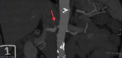
Figure 1:
• Mural thickening/eccentric thrombus of the proximal SMA (Figure 2).
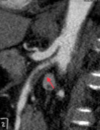
Figure 2:
CT Angiogram Aortic Arch & Carotids
• Severe stenosis of the origin of the left ICA with significant circumferential thickening and perivascular stranding (Figure 3).
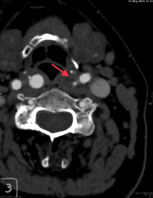
Figure 3:
• Evidence of mild disease at origin of the right ICA (Orange arrow - Figure 4).
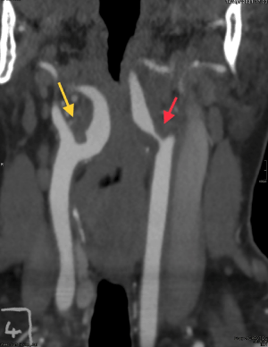
Figure 4: