
Rapid Communication
Austin J Radiol. 2021; 8(12): 1173.
Appropriate Use of Lumbar Spine X-Ray for Low Back Pain-An Investigation of Limited Clinical Value
Saadawy A*
Department of Radiology, West Suffolk Hospital, Addenbrookes Hospitals, UK
*Corresponding author: Ahmed Saadawy, Department of Radiology, West Suffolk Hospital, Addenbrookes Hospitals, UK
Received: October 22, 2021; Accepted: November 17, 2021; Published: November 24, 2021
Introduction
NICE has been advising for years against routine imaging for low back pain, unless the result is likely to change patient management. The recent GIRFT report further highlighted this as an area of concern and that we should be reducing ’low value imaging’.
In 2017-18, >400,000 lumbar X-rays were undertaken across England. Just over half of these from GP referrals.
Back pain is usually the result of conditions that cannot be diagnosed on XR (osteoporotic collapse an exception). MRI if physiotherapy did not help.
Referrals for lumbar spine x-ray from non-specialists are currently rejected in the absence of red flag symptoms and referral to physiotherapy is recommended.
Why Not Just Start With an XR?
431060 - 68 y/o Female
The delay between XR and appropriate investigation: (21st May – 13 July 2021)
XR:
• Good alignment of thoracic and lumbar spine. No fracture.
• Some degenerative changes but no evidence of aggressive disease.
MRI:
• Tumour replacement of the body of L1, T12 with canal narrowing.
• Another MRI done 7 days later showed progression compared to initial MRI.
M05 Acute Back Pain (≤6 Weeks) with Potentially Serious Features
Neurological (cauda equina syndrome)
• Sphincter and gait disturbance (Table 1-3)
![]()
Modality
Dose
Recommendation(Grade)
Comment
MRI
None
Indicated only in specific circumstances[c]
MRI is the preferred investigation for the diagnosis of most spinal diseases and is helpful in identifying those patients who may benefit when planning surgical Intervention or pain management.
XR
Indicated only in specific circumstances[c]
XR is only indicated if presentation suggests osteoporotic collapse in the elderly.
CT
Indicated only in specific circumstances[c]
CT is used when MRI is contraindicated and when further assessment
Of spondylolysis is required.NM(bone scan)
Indicated only in specific circumstances[c]
NM is non-specific and has been largely superseded by MRI and CT in the
Assessment of chronic back pain. It may show occult osteoid osteomas and
Spondylolysis.Lumbar imaging for low back pain without suggestion of serious underlying conditions does not improve clinical outcomes.
Table 1: M04 Chronic lumbar back pain (>6 weeks) with no clinical or serological indicators of infection or neoplasia (i.e., no red flags).
![]()
Modality
Dose
Recommendation
Comment
MRI
None
Indicated [B]
MRI is the imaging investigation of choice.it is
indicated immediately in patients with acute
neurological features and urgently in those with
Suspected malignancy or infection.XR
Indicated only in
specific circumstances[C]Plain radiograph may be required preoperatively
MRI is preferable as the first line investigation in
patients with potentially serious features, since it
Has a stronger negative predictive value.CT
Indicated only in
specific circumstances[C]CT is an alternative to MRI(when not feasible)
is useful to guide soft tissue/bone biopsyNM(bone scan)
Indicated only in
specific circumstancesBone scintigraphy, especially with SPECT, can help
To identify metabolically active lesions. Specificity
will be increased by comparison with XR or co.
Table 2: M05 Acute back pain (≤6 weeks) with potentially serious features.
![]()
Modality
Dose
Recommendation
Comment
MRI
None
Specialised investigation[B]
For patients with non-specific back pain(with no radicular
Symptoms or red flags), MRI does not help clinical outcome.
It should be reserved for patients referred for orthopedic
Opinion. MRI is the preferred investigation( wider field of
View visualising the conus, postoperative changes, etc.).CT
Specialised investigation[B]
CT only if fracture is suspected or MRI not feasible.
XR
Indicated only in specific
circumstances [C]Acute back pain is usually the result of conditions that
cannot be diagnosed on XR(osteoporotic collapse is an
Exception). Normal XR may be falsely reassuring.(Spinal malignancy, infection, fracture, cauda equina syndrome, ankylosing spondylitis or another inflammatory disorder).
Table 3: M06 Acute back pain (=6 weeks) without potentially serious features.
• Saddle anaesthesia
• Severe or progressive motor loss
• Widespread neurological deficit
Other features include
• Previous malignancy
• Immunosuppression
• Steroid use
• Fever
Aim, Objectives & Standards
Aim
To assess the number of x-rays performed inappropriately for low back pain in the absence of red flag symptoms.
Standards
Lumbar spine X-rays performed from GP referral for back pain without red flag symptoms or concern for Fracture (Target = 0%)
Methodology
• Data collected via CRIS search with the help of Amanda Yeldham.
• Lumbar spine x-rays performed via a GP referral.
• January to June 2021 timescale.
• Referral forms evaluated for information present assessing for any red flag symptoms or other acceptable reasons for performing x-ray.
• Data collected into an excel sheet to allow further analysis.
NICE-Red Flags
Spinal fracture
Red flags include:
• Sudden onset of severe central spinal pain which is relieved by lying down.
• A history of major trauma (such as a road traffic collision or fall from a height),
• minor trauma, or even just strenuous lifting in people with osteoporosis or those who use corticosteroids.
• Structural deformity of the spine (such as a step from one vertebra to an adjacent vertebra)may be present.
• there may be point tenderness over a vertebral body.
Cancer
Red flags include:
• The person being 50 years of age or more.
• Gradual onset of symptoms.
• Severe unremitting pain that remains when the person is supine, aching night pain that prevents or disturbs sleep, pain aggravated by straining (for example, at stool, or when coughing or sneezing), and thoracic pain.
• Localised spinal tenderness.
• No symptomatic improvement after four to six weeks of conservative low back pain therapy.
• Unexplained weight loss.
• Past history of cancer-breast, lung, gastrointestinal, prostate, renal, and thyroid cancers are more likely to metastasize to the spine.
Results
See Figure 1.
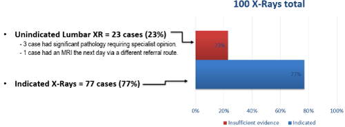
Figure 1:
Limitations
Data collection
The reliance on a request that does not fulfil the criteria can be misleading when the patient has an underlying pathology that is not mentioned, however, would have been an indication for XR.
Data analysis
Implementing the red flags when reading requests can sometimes be difficult when the clinical information is vague/the referrer is a poor communicator.
Action Required, Learning and Improvement
• Radiologists to support radiographers in rejecting referrals for lumbar spine x-rays and directing patients to appropriate pathways. Reminder to radiographers of red flag symptoms/signs.
• Review and amend departmental justification procedures, policies, rules and standards for lumbar radiography.
• Amending the referral form for GPs to be more readable and to contain easier to implement guidance and awareness of red flags.
• Re-audit aiming for 100% compliance with standards.
Acknowledgements
Project lead: Dr Elliott Rees, for his continuous support and encouragement in this project and others.