Clinical Image
Austin J Radiol. 2021; 8(12): 1177.
A Rare Case of Hirayama Disease in a 28-Year-Old Indian Woman
Mathew H* and Ashwin KP
Kasturba Medical College Manipal, India
*Corresponding author: Hannah Mathew, Kasturba Medical College, India
Received: November 10, 2021; Accepted: December 01, 2021; Published: December 08, 2021
Clinical Image
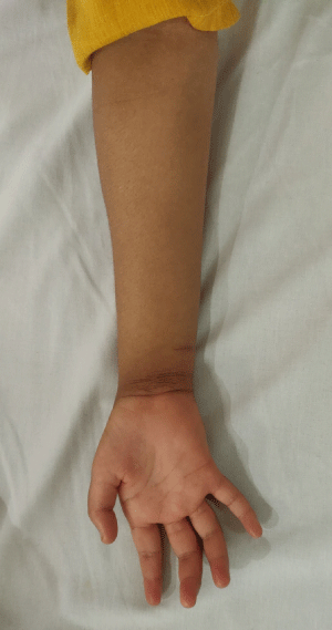
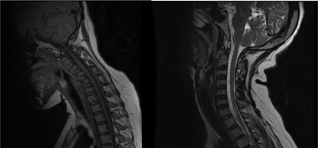
A 28-year-old woman presented with slowly progressive weakness of the right upper limb since 14 years of age. This was purely motor, distally located and progressive in nature. It did not involve any other limbs. She also had a history of hypothyroidism and gestational diabetes. On examination, hypothenar muscle wasting and complete claw hand was noted (Figure 1) Power was reduced in muscles of the hand, while reflexes were bilaterally absent on both upper limbs. Distal asymmetric motor neuropathy was suspected, and nerve conduction studies confirmed the same. Plain MR imaging of the whole spine with dynamic flexion and extension of cervical spine was done which showed features consistent with Hirayama disease (Figure 2).

    Figure 1: Wasting of hypothenar muscles of the right hand and complete
claw hand.

    Figure 2: Craniovertebral junction is normal. Loss of cervical lordosis. On
flexion imaging, there is anterior displacement of posterior dura with enlarged
posterior epidural space (extends from the level of C4 to C7 vertebrae for a
maximum length of ~5.4 cm and maximum thickness of ~4.8mm) showing
epidural flow voids. There is atrophy of lower cervical spinal cord, with
associated intramedullary T2WI/STIR hyperintensity involving the anterior
horn of the spinal cord extending from C4 to C7 vertebrae. At all vertebral
levels, mild diffuse disc bulge indenting the anterior thecal sac was seen, not
resulting in neural foraminal narrowing.
Hirayama Disease (HD) is a rare neurological condition characterized by sporadic juvenile muscular atrophy of the distal upper extremities, which predominantly affects the lower cervical cord. It mainly develops in the late teens and early twenties with a male preponderance ratio of about 20:1. Clinical features include insidious onset and slow progression of unilateral or bilateral muscular atrophy with weakness of the forearms and hands. Sensory disturbance, autonomic involvement, and Upper Motor Neuron (UMN) signs like hyperreflexia and hypertonia are rare. Diagnosis is based on the typical clinical features and dynamic MRI study when the neck is flexed. MR studies in flexion reveal anterior displacement of the posterior wall as well as an enhanced crescent-shaped lesion in the posterior epidural space of the lower cervical canal, which typically disappears when the neck returns to a neutral position. MR imaging studies of the cervical spine in a neutral position can reveal several features such as localised lower cervical cord atrophy, asymmetrical cord flattening, and loss of attachment between the posterior dural sac and subjacent lamina, as well as non-compressed intramedullary high T2 signal intensity.