
Research Article
Austin J Radiol. 2022; 9(1): 1184.
Clinico-Radiological Correlation in COVID-19 Disease: Experience of the Hassan II University Hospital of Fez
Diallo ID*, Sekkat A, Traore WM, Bouardi N, Haloua M, Lamrani MY, Boubou M, Maaroufi M and Alami B²
Department of Radiology, Hassan II University Hospital, Fez, Morocco
*Corresponding author: Diallo Ibrahima Dokal, Service of Radiology, Hospital of Specialties, Avenue Maghreb el araabi, 5 Bis at entrance B Residence, Apt 16.4eme arrondissement, Rabat, Morocco
Received: December 14, 2021; Accepted: January 13, 2022; Published: January 20, 2022
Abstract
Covid-19 disease has a great clinico-radiological polymorphism. Through this study, we illustrate the correlation between these lesions and the clinical presentation of patients infected by SARS-CoV-2 at the Hassan II University Hospital of Fez during the second wave.
Keywords: COVID-19; Correlation; CT scan; Clinical; Fez
Abbreviations
SARS: Severe Acute Respiratory Syndrome; 2019-nCOV2: Related to the New Coronavirus 2019; HR CT: High Resolution Computed Tomography; NSAIDs: Non Steroidal Anti-inflammatory drugs
Introduction
Covid-19 infection has been a major global public health problem since early 2020 [1].
The pandemic started for the first time in China in Wuhan following the isolation of a new virus genetically related to coronaviruses, responsible among other things for a severe acute respiratory syndrome, SARS COV-2 [2]; which has caused a lot of ink to flow from researchers around the world.
The disease linked to covid-19 is characterized by a great clinical polymorphism ranging from an asymptomatic form to an acute respiratory failure. The usual presentation is essentially respiratory signs occurring in a febrile context, including dry cough, dyspnea, flu-like syndrome. Uncommon forms are possible with various symptoms: diarrhea, anosmia, agueusia, nasal congestion, conjunctivitis, skin rashes and even discoloration of the hand or foot [3].
Para-clinical examination remains the cornerstone of the diagnosis and is based on:
• Imaging, particularly thoracic CT, which mainly reveals ground glass opacities and condensation foci characterized by their peripheral distribution.
• But also biology, which allows the final diagnosis to be made thanks to PCR and serological techniques which identify the virus; in almost 80% of cases, the blood count shows lymphopenia [1].
The management of patients is essentially symptomatic; it is based on protocols defined by each country. In Morocco, the Ministry of Health has opted for a treatment combining chloroquine and azithromycin within the hospital [4]; anticoagulation and corticotherapy are an integral part of this management.
Nowadays, vaccination has become the mainstay of the fight against this disease.
The lack of data concerning the subject, the clinico-radiological diversity as well as the prevalence of the infection related to Covid-19, motivated the choice of this topic.
The objective of this study is to determine the relationship between radiological aspects and clinical presentation in patients confirmed Covid-19 positive by PCR.
Materials and Methods
This was a retrospective descriptive and analytical study, conducted at the Hassan II University Hospital in Fez, with 142 patients over a period of 2 months from July 1 to September 1.
Our study included all hospitalized patients who had a positive PCR on nasopharyngeal swab and for whom a CT scan was performed on admission. During this period, all patients with incomplete records or treated on an outpatient basis were excluded from the study. Thus, we divided our patients according to clinical presentation (symptomatic/asymptomatic), severity groups (mild to moderate and severe to critical) and the degree of radiological involvement:
Group 1 (Mild to Moderate) corresponded to patients with pneumonia without signs of severity or mild case with one or more risk factors.
Group 2 (Severe to Critical) included patients with signs of severity requiring intensive care hospitalization with respiratory support.Radiological involvement was assessed according to a score:
• Minimal when the involvement is <10% =score 1
• Moderate when it is between 10 and 25%=score 2
• Extensive for an involvement of 26 to 50%=score 3
• Severe for an involvement between 51 and 75%=score 4
• Critical for > 75%=score 5.
All patients underwent a CT-HR of the thorax, performed in the supine position using the 64-slice Bright Speed scanner (General Electric Medical System, Milwaukee, USA). The acquisitions were made using a slice thickness of 5mm and were reconstructed with a thickness of 1.25mm. Some scans were performed after injection of the contrast medium because of the worsening of the clinical condition, which raised the suspicion of a pulmonary embolism. Chest CT data were reviewed by junior radiologists and approved by senior radiologists.
The chi-square statistical test was used to investigate the relationship between these different parameters with a significance level of 5% (P-value <0.05) using SPSS (statistical package for social sciences) software.
Results
The median age in our series was 59 years with extremes ranging from 20 to 90 years. There was a male predominance (65%, n=93) (Graph 1).
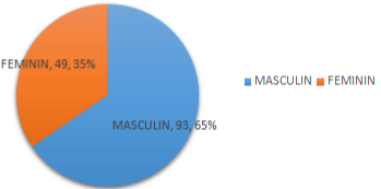
Graph 1: Distribution of patients by gender.
The majority of our patients had defects (58%, n=82). Arterial hypertension, diabetes, and heart disease, which could be associated in the same patient, were the most common defects and concerned mainly symptomatic subjects (Graph 2).
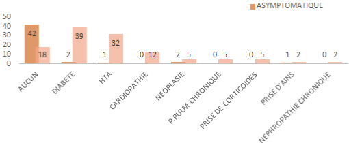
Graph 2: Répartition des antécédents selon la présentation Clinique.
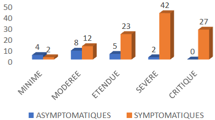
Symptomatic patients were the most represented in our series (79.6%; n=113). The majority of them belonged to clinical severity group 2 (84%; n=95) with febrile respiratory distress in the foreground (45%; n=51) (Graph 3 and 4).
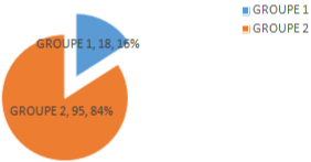
Graph 3: Répartition des patients symptomatiques selon la sévérité Clinique.
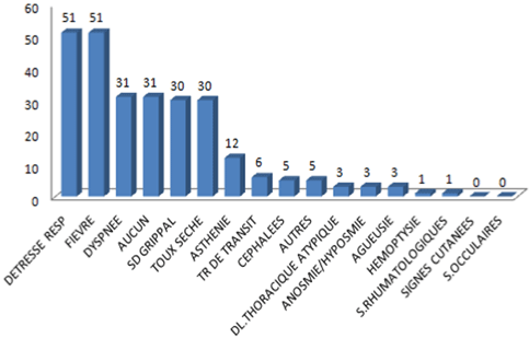
    Graph 4: Representation of the symptoms of our patients according to the
workforce.
The mean time to CT scan was 20 days since the onset of symptoms. Viral pneumonia was present in 88% of cases (n=125 out of 142), mostly in symptomatic subjects in the form of ground glass and condensation foci (Graph 5, Figure 1, Table 1). Several lobes were affected in the majority of cases 67% (n=84), followed by lower lobe involvement 48% (n=60) (graph 6). Peripheral (87%), subpleural (70%) distribution was predominant.
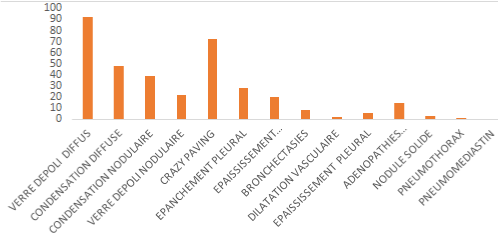
Graph 5: Anomalies scanographiques retrouvées chez nos patients.
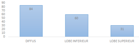
Graph 6: Illustration du siège de l’atteinte scanographique.
    Graph 7: Distribution of patients according to the degree of radiological
severity (P-value <0.001).
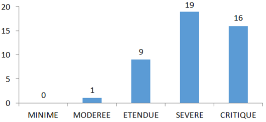
    Graph 8: Bar chart showing the number of deaths according to the degree of
radiological severity (P-value <0.001).
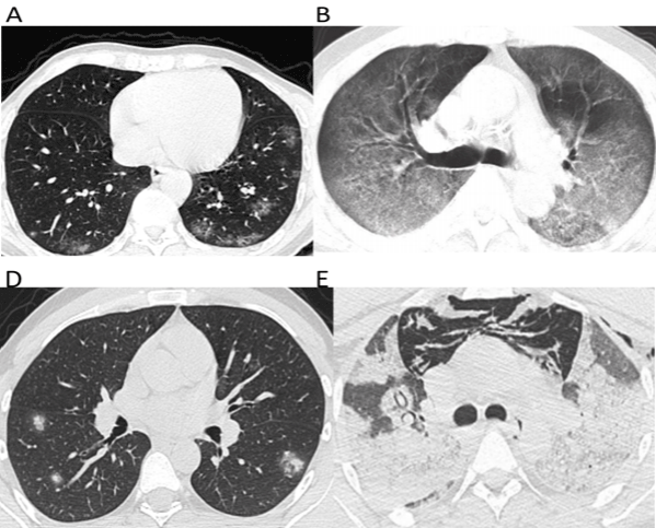
    Figure 1: Density anomalies most commonly encountered in COVID-19.
A) Nodular, patchy ground-glass foci of subpleural, peripheral and
posterobasal topography. B) Diffuse ground-glass foci of mixed topography.
D) Nodular condensation foci of a SARS COV-2 patient, which were
predominantly peripheral, surrounded by ground glass realizing the halo sign.
E) Focuses of diffuse condensation of mixed topography. Note the presence
of pneumomediastinum and subcutaneous emphysema in a patient who
became secondarily comlinked.
The extent of lesions was greater in symptomatic patients than in asymptomatic ones (p <0.001) with a predominance of severe involvement (score=4).
The evolution was unfavorable in some patients with 33% (n=47) of death which was by far in symptomatic patients (n=45). Radiologically, patients with severe to critical involvement (score 4 and 5) had the highest mortality rates among deaths, namely 40.4% (n=19) and 34% (n=16) respectively.
Moderate, extensive and mild forms reached 2% (n=1), 17% (n=8) and 0% respectively. The other 10 patients with moderate to extensive involvement died of metabolic causes (acid ketosis decompensation with disturbance of consciousness, renal), vascular causes (pulmonary embolism in one patient) and particular terrain (cancer, pneumothorax, giant lobar emphysema).
Discussion
Covid-19 disease is a pandemic of viral origin related to SARS COV-2 of the coronaviridae group, responsible for an acute respiratory syndrome. It has attracted the attention of health professionals and scientists all over the world because of its dangerousness in terms of infection power and lethality. A great polymorphism characterizes this disease both clinically and radiologically. Demographically, elderly males are the most likely to develop severe forms of this disease because of the defects and the weakness of the immune response in these patients. In fact, according to retrospective studies conducted in Wuhan hospital, the average age of Covid-19 patients was 55.5 [5] years, 49 [6] years, 56 [7] years, which is comparable to the average age in our study which was 59.5 years. Regarding gender, to date, several studies have addressed the gender disproportion in the incidence of COVID-19 disease. Some studies have shown that women are less likely to acquire viral infections. Further analysis of the underlying causes suggests that they have higher macrophagic and neutrophilic activity and higher antibody production [8].
In addition, in vivo studies on angiotensin-converting enzyme 2 (ACE2), which is the protein involved in the first step of SARS COV- 2 viral entry, have shown a higher expression in the kidneys of male than female patients, which may explain, according to these studies, the male predominance of COVID-19. However, this remains to be demonstrated as it is not clear whether there are gender differences in pulmonary ACE2 receptor expression [8].
On the other hand, a growing number of studies show no statistically significant gender differences in SARS-CoV-2 infections. In our series, there was a predominance of males (65%), as in several studies, including that of Gebhard et al., which showed a male predominance [9].
Clinically, the majority of patients were symptomatic (nearly 80%) with a largely noisy symptomatology, which can be explained by the advanced age of the patients with an average of 64.75 years and their previous history (only 28% had no history) (graph 2). Several studies have pronounced on the main risk factors for the occurrence of severe forms (Huang 2020, Guan 2020); in Wuhan, there was a clear and considerable relationship between age and risk of infection on the one hand (especially with severe forms) and poor prognosis (death) on the other hand (Wu 2020). According to the Italian National Institute of Health, an analysis of the first 2003 cases of death revealed that the median age was 80 years, only 17 (0.8%) were 49 years or younger while 88% were older than 70 years (Livingston 2020) [10].
In addition to advanced age, several comorbidities, such as hypertension, have been identified as major risk factors for severe disease and death. In addition, mild clinical forms benefited from ambulatory management during the study period corresponding to the 2nd wave in Morocco and were therefore not included in our study, which further increases the severity score of our patients during this period.
Respiratory manifestations constitute the main symptom of SARS COV-2 infection and are dominated by dry cough and respiratory difficulty. Many studies conducted in China have reported this (Chen et al. [11]; Guan et al. [12]; Wang [7,13]; Zhang et al. [14]).
Indeed, all these studies highlighted the frequency of dry cough which was present in (43-82%), Respiratory distress in (18-55%), dyspnea in 18.7%, hemoptysis in 1% in patients infected with SARSCOV- 2. These data are comparable to our study with similar results for respiratory distress which was found in 35.9% of cases, dyspnea in 21.8% and hemoptysis in almost 1% of cases; a little less for dry cough which was present in 21%.
Chest CT without injection is the reference examination: it is useful for triaging symptomatic patients who may or may not require hospitalization in isolation in the absence of or while awaiting the results of RT-PCR; it is also useful for differential diagnosis, evaluation of severity and search for complications. Its performance is highly variable depending on the series, with sensitivities ranging from 44 to 98%, while specificity varies between 25 and 94% (on average less than 50%) [15,16]. Sensitivity could increase according to the delay from the onset of symptoms: 84% between days 0 and 5, and 99% between days 6 and 11 [17]. In our study, the sensitivity of the CT scan was 88%, higher in symptomatic patients (94%; n=106). These results are close to those of a Chinese series, covering 211 patients, conducted by Song and colleagues [18], in a hospital in Wuhan, who had 205 positive scans (sensitivity of 97%), of which 55 (26%) were diagnosed by chest CT with negative RT-PCR.
In our facility, the indications for CT scans have evolved according to the epidemiological situation, the resources available and the organizational aspects of the hospital. Thus, initially during the containment period, CT scanning was indicated for all covid-19 patients, as long as most of the hospital’s activity was devoted to this pandemic, but with the evolution of the epidemiological situation, and in particular after decontainment and the resumption of various hospital activities, the indication of the CT scan became reserved to symptomatic cases especially with comorbidity or desaturation, as well as to patients who presented to the emergency room in respiratory distress before the PCR test was carried out, in order to decide on the route that they would take.
The frequencies of radiological signs noted in our study (Graph 5) are similar to those observed in the literature. In fact, in similar studies [19,20], the frequencies of the most common radiological signs were variable: ground glass (86-91%), condensation focus (63%); the correlation between these lesions and clinical severity has been demonstrated by several publications, such as a Chinese study carried out in Jiangsu province (p<0.001). In this study, ground glass was more present in patients with moderate to severe symptoms as well as condensation compared to those with mild symptoms [21].
In our study, no significant association was found between the occurrence of these lesions and the clinical presentation (symptomatic or not) of patients infected with COVID-19 (Table 1).
![]()
    Ground Glass
    Condensation


    Asymptomatic
    21(18.4%)
    17(19.5%)


    Symptomatic
    93(81.6%)
    70(80.45%)


    P-value
    0.502
    0.361
Table 1: Distribution of the most frequent anomalies in our series.
The peripheral sub pleural topographic distribution found in our study is similar to that described in several publications with peripheral (75%), multi lobar (89%), posterior topography (80%) and predilection for the lower lobes [22,23].
The main CT sign of severity is the extent of parenchymal abnormalities. Numerous studies report a correlation between the extent of lesions and clinical severity [24-26]. Indeed, K Li et al. have noted a strong correlation between the degree of radiological severity and the clinical severity of symptoms in patients infected with SARSCOV- 2 (Table 2). Thus, in a comparative study conducted in China on 25 patients with severe to critical clinical presentation and 58 others with benign symptomatology, qualified as ordinary patients in this study, it was shown that the scanographic scores of the severe/ critical patients were significantly higher than those of the ordinary patients (11 vs. 5) (P <0.001). On the other hand R .Yang et al., had found that the individual scores in each lung and the total score were higher in severe COVID-19 compared with mild cases (P <0.05) [26].
![]()
Imagery
SymptomsGround Glass
Condensation
Asymptomatic/leger (n=63; 13%)
0.1
0.4
Moderate (n=378; 78.1%)
9.1
97.5
Grave (n=43; 8.9%)
14.8
437.3
P-value
<0.001
<0.001
Table 2: Table, illustrating the correlation between the appearance of the most
frequent lesions and clinical severity in covid-19 patients in Jiangsu Province,
China [31].
In line with this literature data, our study perfectly illustrates this radio-clinical correlation; indeed, more than half of the symptomatic patients who belonged to group 2, had the highest radiological severity scores (between 4 and 5) whereas the asymptomatic patients had mostly a minimal score (p <0.001). The severity of the scanographic involvement could be explained by the long interval between the onset of symptoms and the thoracic scan. Indeed, all these patients had a CT scan delayed from the onset of symptoms (with a mean interval of 20 days). In addition, these patients had several risk factors, such as:
• High age: severe forms were associated with an age >50 years which is consistent with the study of k Li et al.
• Male sex: 60% of patients who developed severe forms were men.
• Hypertension, diabetes, anti-inflammatory drugs, lymphopenia and elevated C-reactive protein are incriminated as risk factors for severe disease.
The establishment of this radio-clinical correlation is of major interest in determining the prognosis of the patients and thus in their orientation towards the most appropriate COVID-19 management structures.
Some teams use other severity criteria, in particular parenchymal density (quantified by scintigraphy), pleural effusions and early architectural deformities [27]. A Chinese series by K li et al. also suggests that an initial involvement of the upper lobes may be a marker of poor prognosis [13]. Indeed, this study reported that right upper lobe involvement was associated with severe forms of covid-19 (odd ratio 5.603; p=0.029). Some studies have established severity prediction scores, in the light of a Dutch study, conducted by Schalekamp and colleagues, which established a risk calculator, made available for download [28] (Table 3 and 4), according to gender, comorbidities, clinic, radiological findings.
![]()
    Variables
    Description


    Male sex
    Male sex


    Obstructive lung disease
    Medical diagnosis of asthma or chronic obstructive    pulmonary disease


    > 7 days complaints
    Self-reported symptoms for more than 7 days on hospital    admission


    Neutrophils (109/l)
    Blood neutrophil level


    C-reactive proteine (mg/l)
    Blood C-reactive proteine level


    LDH (IU/l)
    Blood lactate deshydrogenase level


    Distribution
    Distribution pattern on the chest x-ray, either peripheral    diffuse, central or normal


    Chest x-ray score
    Value 0 to 8.sum of score per quadrant: no involvement    (>50%)=2 points.
Table 3: Dutch prognosis calculator for COVID-19.
![]()
    Variables
    Select input below
    Equivalent value


    Male sex
    yes
    1


    Obstructive lung disease
    no
    0


    > 7 days complaints
    yes
    1


    Neutrophils (109/l)
    6.1
    6.1


    C-reactive protein (mg/l)
    177
    177


    LDH (IU/l)
    302
    302


    Distribution
    Peripheral
    0


    Chest x-ray score
    6
    6


    Risk of USI/Death %
    39.2
Table 4: Example of a Dutch prognosis calculator for COVID-19.
A part from clinical factors, radiology can give an indication of the evolution of the patients thanks to a certain number of parameters, namely the quantification of the extension of the lesions via a severity score, the diameter of the pulmonary artery and sometimes the patterns of the CT abnormalities and the lesion associations [29].
M Raoufi et al., evaluated 380 patients with a mean age of 53.62 ± 16.66 years (66.1% male). The most frequent chest CT abnormalities were peripheral (86.6%) and peribronchovascular (34.6%), with a ground glass pattern (54.1%) and a round (53.6%) or linear (46.7%) shape. There was a significant correlation between the shape of the abnormalities (p = 0.003), the CT severity score (CTSS) (p <0.0001), the diameter of the pulmonary artery (p = 0.01) and mortality. The mean radiological severity score of the non-surviving cases was significantly higher (13.68 ± 4.59 vs. 8.72 ± 4.42; <0.0001), which is consistent with the results of our study. In our study, patients with a radiological score of 4 to 5 had the highest mortality rates, ranging from 45% and 57% respectively, followed by patients with a score of 3 (27% mortality) (p<0.0001). It is therefore necessary to remain vigilant in the presence of moderate CT damage, especially if there are associated defects (Table 5).
![]()
    Settings
    Effective    (n=83)
    Severe/    Critique (n=25)
    Ordinary    (n=58)
    p-value


    CT score
    5 (4-8)
    11 (8-15.5)
    5 (2.5-5)
    <0.001


    Ground glass
    81 (97.6%)
    25 (100.0%)
    56(96.6%)
    1.000


    Condensation
    53(63.9%)
    22(88%)
    31(53.4%)
    0.001


    Linear opacities
    54(65.1%)
    23(92%)
    31(53.4%)
    0.003


    Interlobular septal thickening
    52(62.7%)
    19(76%)
    33(56.9%)
    0.099


    Crazy paving
    30(36.1%)
    16(64%)
    16(27.6%)
    0.013


    Sub pleural curvilinear line
    17(20.5%)
    8(32%)
    9(15.5%)
    0.088


    nodules
    6(7.2%)
    3(12%)
    3(5.2%)
    <0.001


    reticulations
    4(4.8%)
    3(12%)
    1(1.7%)
    0.079


    Pleural effusion
    7(8.4%)
    7(28%)
    0(0%)
    <0.001


    Pericardial effusion
    4(4.8%)
    7(28%)
    0(0%)
    <0.001


    lymphadenopathy
    7(8.4%)
    4(16%)
    0(0%)
    0.007


    Right upper lobe
    62(64.7%)
    23(92%)
    39(67.2%)
    0.017


    Right middle lobe
    61(73.5%)
    22(88%)
    39(67.2%)
    0.049


    Right lower lobe
    78(94%)
    25(100%)
    53(91.4%)
    0.316


    Upper Lobe Left
    71(85.5%)
    24(96%)
    47(81.0%)
    0.150


    Lower Lobe Left
    80(96.4%)
    25(100%)
    55(94.8%)
    0.550


    Upper lobes
    74(89.2%)
    24(96%)
    50(86.2%)
    0.316


    Lower lobes
    80(96.4%)
    25(100%)
    55(94.8)
    0.550


    bilaterality
    79(95.2%)
    25(100%)
    54(93.1%)
    0.310


    Neither lobe affected
    5(4-5)
    5(5-5)
    5(3-5)
    0.003
Table 5: Results of a study illustrating the radio-clinical correlation in covid-19
patients in the study by Li et al. [32].
On the other hand, the existence of a particular clinical form known as silent hypoxia, which is fortunate [30], where patients with severe damage on the CT scan do not present any symptoms or at least remain minimally symptomatic, makes CT essential in the management of covid-19 patients.
The majority of these deaths were related to respiratory distress due to pulmonary extension of the lesions caused by COVID-19 responsible for deep desaturation.
However, some deaths were related to the extension of the lesions, to a particular terrain or following a complication, thus we noted a case of pulmonary embolism, 1 case of pulmonary cancer, 1 case of bilateral apical centrilobular emphysema with pneumomediastinum, 1 case of panlobular emphysema and 1 case of pneumothorax. In addition although not having very extensive lesions, some patients died as a result of metabolic problems (3 patients in acid-ketotic decompensation with consciousness disorder) or organ dysfunctions; this is the case of 2 patients who presented cardiac rhythm disorders, one of which on acute coronary syndrome, also of a patient in terminal renal failure and three other patients who were in consciousness disorder on stroke.
Conclusion
The SARS COV-2 infection is a disease with variable manifestation, mainly affecting the lung, however a multi-systemic involvement remains very possible and potentially formidable.
The diagnosis of certainty of this infection is possible thanks to PCR. However, HR chest CT has become essential in the management of COVID-19 patients. Indeed, it offers good sensitivity, recognizing aspects suggestive of COVID-19 pneumonia including density abnormalities, made of frosted glass foci and condensations of peripheral topography, sub pleural, predominantly posterobasal, which allows to refer patients to a COVID-19 circuit, thus avoiding prolonged contact with these patients.
It also allows to eliminate other differential diagnoses that may be confused with COVID-19 pneumonia. Another major role of the CT scan is to assess the degree of radiological severity of the disease, which is strongly correlated with the clinical severity and thus plays a role in the prognostic evaluation of patients. This was demonstrated in our study, where we noted a poor prognosis in patients with severe to critical lung parenchymal damage on CT, particularly in elderly multi-targeted subjects.
A study on a large sample would be desirable in order to reinforce these results which remain limited by the size of the sample studied and its retrospective character.
References
- Tsang HF, Chan LWC, Cho WCS, Yu ACS, Yim AKY, Chan AKC, et al. An update on COVID-19 pandemic: the epidemiology, pathogenesis, prevention and treatment strategies. Expert Rev Anti Infect Ther. 2021; 19: 877-888.
- Zhu N, Zhang D, Wang W, Li X, Yang B, Song J, et al; China Novel Coronavirus Investigating and Research Team. A Novel Coronavirus from Patients with Pneumonia in China, 2019. N Engl J Med. 2020; 382: 727-733.
- Lipsker D. Paraviral eruptions in the era of COVID-19: Do some skin manifestations point to a natural resistance to SARS-CoV-2? Clin Dermatol. 2020; 38: 757-761.
- Ministère de la santé Maroc. Covid-19 [en ligne]. 2020.
- Chen N, Zhou M, Dong X, Qu J, Gong F, Han Y, et al. Epidemiological and clinical characteristics of 99 cases of 2019 novel coronavirus pneumonia in wuhan, China: a descriptive study. The Lancet. 2020e: 395: 507-513.
- Huang C, Wang Y, Li X, Ren L, Zhao J, Hu Y, et al. Clinical futures of patients infected with 2019 novel coronavirus in wuhan, China. The Lancet. 2020a; 395: 497-506.
- Wang D, Hu B, Hu C, Zhu F, Liu X, Zhang J, et al. Clinical Characteristics of 138 Hospitalized Patients With 2019 Novel Coronavirus-Infected Pneumonia in Wuhan, China. JAMA. 2020; 323: 1061-1069.
- Kopel J, Perisetti A, Roghani A, Aziz M, Gajendran M, Goyal H. Racial and Gender-Based Differences in COVID-19. Front Public Health. 2020; 8: 418.
- Gebhard C, Regitz-Zagrosek V, Neuhauser HK, Morgan R, Klein SL. Impact of sex and gender on COVID-19 outcomes in Europe. Biol Sex Differ. 2020; 11: 29.
- B Sebastian K, C hoffmann et al. Covid reference. ENG 2020/05.
- Chen N, Zhou M, Dong X, Qu J, Gong F, Han Y, et al. Epidemiological and clinical characteristics of 99 cases of 2019 novel coronavirus pneumonia in wuhan, China: A descriptive study. The lancet 2020e: 395: 507-513.
- Guan WJ, Ni ZY, Hu Y, Liang WH, Ou CQ, He JX, et al; China Medical Treatment Expert Group for Covid-19. Clinical Characteristics of Coronavirus Disease 2019 in China. N Engl J Med. 2020; 382: 1708-1720.
- Li LQ, Huang T, Wang YQ, Wang ZP, Liang Y, Huang TB, et al. COVID-19 patients’ clinical characteristics, discharge rate, and fatality rate of metaanalysis. J Med Virol. 2020; 92: 577-583.
- Tang C, Zhang K, Wang W, Pei Z, Liu Z, Yuan P, et al. Clinical characteristics of 20,662 patients with COVID-19 in mainland China: A systemic Review and meta-analysis. medRxiv. 2020.
- Ai T, Yang Z, Hou H, Zhan C, Chen C, Lv W, et al. Correlation of Chest CT and RT-PCR Testing in Coronavirus Disease-2019 (COVID-19) in China: A Report of 1014 Cases. Radiology. 2020: 200642.
- Dangis A, Gieraerts C, De Bruecker Y, Janssen L, Valgaeren H, Obbels D, et al. Accuracy and reproducibility of low-dose submillisievert chest CT for the diagnosis of COVID-19. Radiology: Cardiothoracic Imaging. 2020; 2.
- Wang Y, Dong C, Hu Y, Li C, Ren Q, Zhang X, et al. Temporal Changes of CT Findings in 90 Patients with COVID-19 Pneumonia: A Longitudinal Study. Radiology. 2020: 200843.
- S Song, F Wu, Y Liu, H Jiang, F Xiong, X Guo, et al. Correlation between chest CT findings and clinical features of 211 COVID 19 suspected patients in Wuhan, China. Open forum infectious diseases. 2020; 7: 171.
- Wu J, Wu X, Zeng W, Guo D, Fang Z, Chen L, et al. Chest CT Findings in Patients With Coronavirus Disease 2019 and Its Relationship With Clinical Features. Invest Radiol. 2020; 55: 257-261.
- Zhao W, Zhong Z, Xie X, Yu Q, Liu J. Relation Between Chest CT Findings and Clinical Conditions of Coronavirus Disease (COVID-19) Pneumonia: A Multicenter Study. AJR Am J Roentgenol. 2020; 214: 1072-1077.
- Wang YC, Luo H, Liu S, Huang S, Zhou Z, Yu Q, et al. Dynamic evolution of COVID-19 on chest computed tomography: experience from Jiangsu Province of China. Eur Radiol. 2020; 30: 6194-6203.
- Salehi S, Abedi A, Balakrishnan S, Gholamrezanezhad A. Coronavirus disease2019 (COVID-19) imaging reporting and data system (COVID-RADS) and common lexicon: a proposal based on the imaging data of 37 studies. 2020: 1-13.
- Ye Z, Zhang Y, Wang Y, Huang Z, Song B. Chest CT manifestations of new coronavirus disease 2019 (COVID-19): a pictorial review. Eur Radiol. 2020: 1-9.
- Xiong Y, Sun D, Liu Y, Fan Y, Zhao L, Li X. Clinical and High-Resolution CT Features of the COVID-19 Infection: Comparison of the Initial and Follow-up Changes. Invest Radiol. 2020; 55: 332-339.
- Li K, Wu J, Wu F, Guo D, Chen L, Fang Z. The Clinical and Chest CT Features Associated with Severe and Critical COVID-19 Pneumonia. Invest Radiol. 2020; 55: 327-331.
- Yang R, Li X, Liu H, Zhen Y, Zhang X, Xiong Q. Chest CT Severity Score: An Imaging Tool for Assessing Severe COVID-19. Radiol Cardiothoracic Imaging. 2020; 2: e200047.
- Shi H, Han X, Jiang N, Cao Y, Alwalid O, Gu J, et al. Radiological findings from 81 patients with COVID-19 pneumonia in Wuhan, China: a descriptive study. Lancet Infect Dis. 2020; 20: 425-434.
- Dutch COVID 19 prognostic model.
- Raoufi M, Safavi Naini SAA, Azizan Z, Jafar Zade F, Shojaeian F, Ghanbari Boroujeni M, et al. Correlation between Chest Computed Tomography Scan Findings and Mortality of COVID-19 Cases; a Cross sectional Study. Arch Acad Emerg Med. 2020; 8: e57.
- Couzin-Frankel J. The mystery of the pandemic’s ‘happy hypoxia’. Science. 2020; 368: 455-456.