
Case Report
Austin J Radiol. 2022; 9(1): 1185.
Bifocal Squamous Cell Carcinoma (Bladder and Renal Pelvis): A Case Report
Mejri R¹*, Mrad Dali K², Chaker K², Ben Rhouma S², Ouanes Y² and Nouira Y²
¹Departement of Urology, Hospital Mongi Slim La Marsa, Tunisia
²Departement of Urology, La Rabta Hospital, Tunisia
*Corresponding author: Ramzi Mejri, Department of Urology, Hospital Mongi Slim La Marsa, Tunisia
Received: January 12, 2022; Accepted: February 02, 2022; Published: February 09, 2022
Abstract
Urinary squamous cell carcinoma is a rare variety of urothelial tumors. We report a case of squamous cell carcinoma with dual location in the bladder and renal pelvis resulting in a fatal outcome in a patient suffering from a history of tobacco intoxication and recurrent upper urinary tract infections due to kidney stones. The CT scan showed a bladder tumor mass and a small left kidney destroyed by lithiasis. The study of the kidney surgical specimen confirmed a second location of squamous cell carcinoma. After a review of the literature, this is the first case of a bifocal squamous cell carcinoma.
Keywords: Squamous cell carcinoma; Renal pelvis; Bladder; Surgery
Introduction
Squamous cell carcinomas of the urothelium are rare histopathological entities, especially in occidental countries. Whatever their location within the urothelium, their prognosis is usually poor.
Given the paucity of published studies on cohorts of patients with bilharzia, little published scientific data is currently available to optimize management. Squamous cell carcinoma is a tumor with a poor prognosis, often discovered at an advanced stage. Radical treatment is immediately indicated.
Bladder cancer is the second most common urological cancer after prostate cancer and is a significant public health problem [1], with a worldwide incidence of 900,000 new cases. We report a case of bifocal bladder and renal pelvis tumor
Case Presentation
A 23-year-old man, a heavy smoker who had not stopped smoking, presented to our urological surgery unit complaining of left low back pain with total hematuria assessing for 6 months. The patient also reported irritative lower urinary tract symptoms such as urinary frequency, burning and urgency. The patient’s medical history included well-controlled diabetes mellitus and reports of repeated episodes of uninvestigated urinary tract infection for several months. His clinical examination showed sensitivity in the left lumbar fossa. The rectal examination revealed a flat, painless prostate with a flexible and mobile bladder floor. However, the urine was slightly hematic and the hemodynamic constants were stable. Her complete blood count showed hyperleukocytosis at 10600/mm3, ferric anemia with hemoglobin at 10.3gm/dl, a moderate inflammatory syndrome (CRP 38mg/l) and a normal coagulation panel. Cytobacteriological analysis of urine was normal as well as his renal function.
In view of this clinical presentation of low back pain with total hematuria and in order to support the diagnosis, an intravenous urography was performed. It showed a small, multi-lithiasis left kidney with a lacunar image of the left bladder horn (Figure 1).
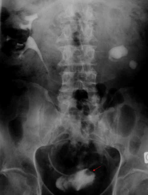
    Figure 1: Intravenous urography: Small lithiasis of the left kidney and lacunar
image of the left horn.
In order to map the bladder lesions, urethrocystoscopy was performed under spinal anesthesia revealing a solid tumor in the left horn of the bladder measuring 2cm in diameter.
Prior to any therapeutic decision, a CT scan was performed showing a small multilithiasic left kidney with laminated parenchyma. There was a large renal pelvis lithiasis piece of 4cm with another lower caliceal lithiasis fragment of 1cm (Figure 2a-2c).
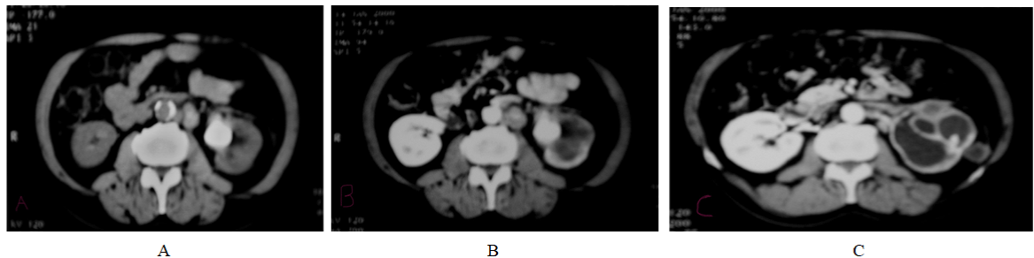
Figure 2: CT scan: A, B) Small left kidney with several lithiasis locations; C) Kidney parenchyma laminated.
The patient underwent endoscopic resection of the bladder tumor and left nephrectomy. Pathological examination found two tumor locations of squamous cell carcinoma: renal pelvis and bladder. A cystoprostatectomy with a left ureterectomy was performed. The evolution was marked by an alteration of the general condition with the appearance of a troisier lymph node at two months follow-up. The patient died of septic shock with a urinary origin and multivisceral failure.
Discussion
Squamous cell carcinoma is an extremely rare tumor in Western countries with a frequency of around 2%, second only to urothelial carcinoma. However, this tumor is much more common in countries of the Middle East and South America where Bilharzia is endemic with a frequency of 20-30%. According to MUSTOFI, the pathophysiological mechanism incriminated in the genesis of squamous cell carcinoma is a response of the bladder epithelium to environmental factors and bladder irritants. This mechanism passes through 3 stages: cell proliferation, metaplasia and neoplasia [2]. Squamous cell carcinoma is a rare histological variety of bladder tumors: 5-8% [3]. Its location in the renal pelvis is still rare. It represents 0.5 to 0.8% of malignant renal tumors. It is believed to occur more frequently in the black population, where the rate of occurrence is approximately 20% of bladder cancers.
The age of discovery of the tumor does not present any particularity compared to other tumors of the urinary tract. A male predominance has been reported by most of the studies.
Tobacco, bilharzia, urinary lithiasis, neurological bladders, radiotherapy, chronic urinary tract infections, the presence of intravesical foreign bodies and any other factor irritating the urothelial mucosa are incriminated in the genesis of this tumor with a significant association [4].
Clinical symptoms are usually non-specific and often dominated by hematuria and atypical back pain, which lose their alarm value because they are often associated with previous infectious episodes. Radiologically, the presence of stones should not delay the positive diagnosis. The association of renal calculi, lacunae and/or a renal mass syndrome with the radiological findings should raise a warning, especially in the case of old calculi. The CT scan remains important for the diagnosis and the assessment of local and distant extension. It is also used to guide biopsies.
The tumor is often infiltrative at the time of diagnosis. This type of cancer is characterized by a mainly local evolution, with less metastatic extension to lymph nodes and visceral organs than in transitional cell carcinomas.
The presence of multiple foci of squamous cell carcinoma in the bladder or upper excretory tract is classic, but the association of upper The presence of multiple foci of squamous cell carcinoma in the bladder or upper excretory tract is classic, but the association of upper
This association seems to worsen the prognosis and poses problems of therapeutic management.
The treatment is surgical, other therapies (partial cystectomy, radiotherapy, chemotherapy) have not proven to be effective [5]. Radical cystectomy combined with pelvic lymph node dissection and urinary diversion is the treatment of choice for bladder squamous cell carcinoma. For locations in the renal pelvis or ureter, an enlarged nephrectomy with total ureterectomy is recommended. In our case of a bifocal lesion, we had combined both types of recommendations. The prognosis of these tumors remains however poor with an average survival of seven months and a five-year survival of no more than 10%.
Conclusion
Urinary squamous cell carcinoma is a rare tumor, which remains unrecognized in Western countries, unlike in North African countries. If the risk factors are well identified, the diagnosis remains late.
Although the risk factors are well identified, diagnosis remains late, which explains the invasive and aggressive nature of this disease at diagnosis. Treatment is surgical. With this type of cancer, prevention becomes very important by treating kidney stones and preventing recurrent upper urinary tract infections.
Authors Contributions
Mejri R: Participated in the writing of the manuscript; Mrad Dali K: Participated in the writing of the manuscript; Chaker K: Participated in the writing of the manuscript; Ben Rhouma S: Participated in the writing of the manuscript and its correction; Ouanes Y: Participated in the writing of the manuscript and its correction; Nouira Y: Participated in the writing of the manuscript and its correction.
References
- Klotz L, Brausi MA. World Urologic Oncology Federation Bladder Cancer Prevention Program: a global initiative. Urol Oncol. 2015; 33: 25-29.
- Rundle J, Hart A, McGeorge A, Smith J, Malcolm A, Smith P. Squamous cell carcinoma of bladder: review of 114 patients. BJU International. 1982; 54: 522-526.
- A Desgrippes, P Meria, A Cortesse, B Cochand-Priollet, G Cariou. Epidermoid carcinoma of the bladder. Prog Urol. 1998; 8: 1097.
- Berry A, Iriart X, Fillaux J, Magnaval J-F. Schistosomose urogénitale et cancer. Bulletin de la Société de pathologie exotique. 2017; 110: 68-77.
- Martin JW, Carballido EM, Ahmed A, Farhan B, Dutta R, Smith C, et al. Squamous cell carcinoma of the urinary bladder: systematic review of clinical characteristics and therapeutic approaches. Arab journal of urology. 2016; 14: 183-191.