Clinical Image
Austin J Radiol. 2022; 9(1): 1186.
The Celery Stalk Sign: An Indicator of Mucoid Cyst of Anterior Cruciate Ligament
Amal L* and En-Nafaa I
Department of Radiology, Mohammed V Military Teaching Hospital, Faculty of Medicine and Pharmacy, Mohammed V University, Rabat, Morocco
*Corresponding author: Lahfidi Amal, Department of Radiology, Mohammed V Military Teaching Hospital, Faculty of Medicine and Pharmacy, Mohammed V University, Rabat, Morocco
Received: January 28, 2022; Accepted: February 21, 2022; Published: February 28, 2022
Keywords
Cystic lesion; Cruciate ligaments; IRM
Clinical Image
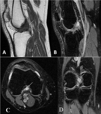
Mucoid cyst of the anterior cruciate ligament is a rare condition that is often overlooked [1,2]. It is a newly formed cystic formation consisting of one or more cavities containing very viscous mucoid fluid and surrounded by a fibro conjunctive wall [2], described by Bergin et al as an uncommon form of mucoid degeneration [1]. Its prevalence in magnetic resonance imaging (MRI) is 0.4 to 1.3% with a male predominance and an average age between 40 and 50 years old [1]. The clinical picture at the beginning is asymptomatic, the evolution is characterized by gonalgia of progressive intensity often posterior in the popliteal fossa [1,2], associated with a limitation of flexion and/or extension with sometimes episodes blocking [1,2]. The physical examination is poor and not very specific, a history of acute trauma or repetitive strain injuries are often involved [1,2]. Magnetic resonance imaging (MRI) is the gold standard for diagnosis [1,2]. It appears in the form of a cystic lesion, fusiform of liquid signal, filling the intercondylar notch and oriented in the long axis of the anterior cruciate ligament which appears widened in a fan “like a stalk of celery” [1,2]. The evolution can be marked by the rupture of the cyst [1] hence the importance of early diagnosis and adequate management. Arthroscopic resection is the treatment of choice given its minimally invasive nature, its good functional results and a nearzero recurrence rate [1,2].

    Figure 1: IRM du genou montre une formation kystique au niveau de la
tente des ligaments croisés en hypo signal T1 (A) et hyper signal T2 franc
(B) sur les coupes sagittales réalisant l’aspect de tige de céleri, comblant
l’échancrure inter condylienne sur les coupes T2 axiale (C) et coronale (D).
References
- M Millet-Luft, S Lovi b, R Rousseau. Les lésions kystiques mucoïdes de la tente des croisés. Journal de Traumatologie du Sport. 2019; 36: 261-264.
- Louaste Jamal, Taoufik Cherrad, Hicham Bousbaa, Hassan Zejjari, Mohammed Ouahidi, Larbi Amhajji, et al. Kyste mucoïde du ligament croisé antérieur: à propos d’un cas. Pan African Medical Journal. 2016; 24: 331.