Case Report
Austin J Radiol. 2022; 9(2): 1189.
Case Report of CT Findings in Post Partum Renal Cortical Necrosis
Neha S¹, Gaurav RA¹, Ankur L² and Ankur S¹*
¹Department of Radiodiagnosis, Dr. Ram Manohar Lohia Institute of Medical Science, Lucknow, India
²Department of Skin and Venerology, VMMC Safdarjung, New Delhi, India
*Corresponding author: Sah Ankur, Department of Radiodiagnosis, Dr. Ram Manohar Lohia Institute of Medical Science, Lucknow, India
Received: February 28, 2022; Accepted: March 19, 2022; Published: March 26, 2022
Abstract
Renal cortical necrosis is ischemic destruction of renal cortex which can be patchy or diffuse caused by significantly reduced arterial perfusion. In addition, direct endothelial injury particularly in setting of sepsis, eclampsia, haemolytic uremic syndrome (HUS) and snake bite may lead to renal ischemia caused by endovascular thrombosis. Progression to end stage renal disease is a rule in diffuse cortical necrosis [1].
Keywords: Cortical necrosis; Post partum; Ischemia; Pregnancy; Acute kidney injury
Introduction
Incidence of acute kidney injury due to renal cortical necrosis is higher in developing countries ranging between 6-7% of all causes of acute renal failure. Complications related to pregnancy are most common cause of renal cortical necrosis. Total ischemic necrosis of all the elements (glomeruli, blood vessels and tubule) of the affected area of the renal cortex secondary to significantly reduced renal perfusion makes it a fatal disease [1].
Case Presentation
A 19-year-old female who was apparently asymptomatic 1-month back when she underwent LSCS for her first pregnancy at 9-month. Following 4 hours after LSCS she developed seizure, for which she was treated. Two days after LSCS there was gradual decrease in urine output and got completely anuric few days after LSCS, with generalized body edema and developed shortness of breath. For the above complaints she underwent hemodialysis and fresh frozen plasma was transfused following which her edema subsided. Following hemodialysis, she developed new onset fever with chills (Figure 1 and 2).

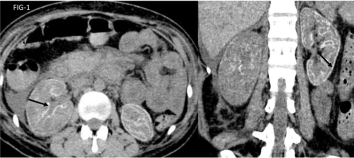
Figure 1: NCCT axial and coronal images showing thin rim of dystrophic cortical calcification (cortical nephrocalcinosis).

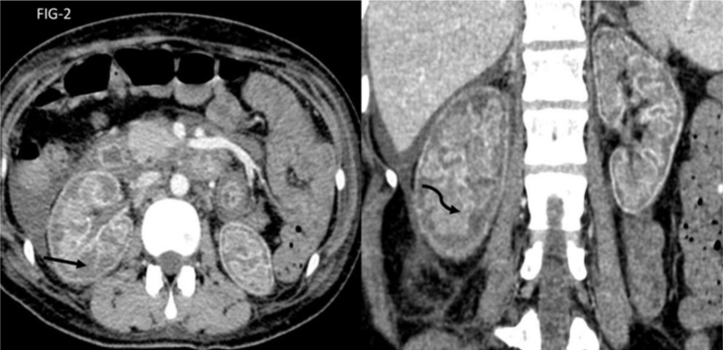
    Figure 2: CECT axial and coronal images showing normal enhancing renal medulla (curved black arrow) with non-enhancing renal cortex (straight black arrow)
giving reverse rim sign.
Investigations
Patient underwent routine blood investigation, blood culture and urine output at time of admission:
Serum urea: 278
Serum creatinine: 14
TLC: 14000
Blood culture was positive for klebsiella and staphylococcus hominis bacteria.
Urine output at time of admission-<100ml per day
Patient underwent CECT for the above complaints after taking consent for use of contrast. Imaging findings are as described.
Discussion
The causes of renal cortical necrosis includes -obstetrical (septic abortion, abruptio placentae, puerperal sepsis, eclampsia, obstetric hemorrhage, intrauterine death and thrombotic microangiopathy of pregnancy) [2] and non-obstetrical (extensive burn, sepsis, HUS, pancreatitis, snake bite, infancy and childhood dehydration, massive GI hemorrhage and diabetic ketoacidosis). Overall obstetric cause is the dominant cause of renal tubular acidosis for 56-61% of cases.
Significantly reduced arterial perfusion is the final common pathway resulting in ischemic necrosis of renal cortex. The vasculature in pregnancy is more sensitive to vasoconstrictors, possibly related to sex hormone [3].
Anuria is the usual presenting symptom of acute RCN. Prolonged anuria in the clinical setting of haemorrhage, sepsis, shock or disseminated intravascular coagulation likely suggests the clinical diagnosis of RCN. The renal biopsy is gold standard for the diagnosis.
USG is the first examination carried out in acute renal cortical necrosis which shows enlarged kidneys and loss of corticomedullary junction [4].
CECT is suitable available non invasive modality for diagnosing RCN. The presence of hypo attenuated subcapsular rim of renal cortex and normal enhancing renal medulla (‘reverse rim sign’) is seen on CECT scan. Due to presence of collateral flow a very thin rim of contrast enhancement (cortical rim sign) may persist which should not be mistaken for adequate perfusion. Eventually dystrophic calcification of the renal cortex may be seen (cortical nephrocalcinosis) [5].
Low signal intensity on both T1 and T2 sequences affecting the renal cortex and column of bertini is the major characteristic finding on MRI [6].
In our report bilateral kidney was diffusely enlarged in size and diffusely hypodense with thin linear area of calcification (black arrows) surrounding renal cortex and around medulla along renal column of bertini (“cortical nephrocalcinosis”). In post contrast images there is evidence of enhancing renal cortex (black straight arrow) and normal enhancing renal medulla giving reverse rim sign (black curved arrow).
Acknowledgements
I would like to acknowledge all my co-authors, who have actively participated in my case report.
References
- Prakash J, Singh VP. Changing picture of renal cortical necrosis in acute kidney injury in developing country. World J Nephrol. 2015; 4: 480-486.
- Smith K, Browne JC, Shackman R, Wrong OM. Acute Renal Failure of Obstetric Origin: An Analysis of 70 Patients. Lancet. 1965; 2: 351-354.
- Chugh KS, Singhal PC, Sharma BK, Pal Y, Mathew MT, Dhall K, et al. Acute renal failure of obstetric origin. Obstet Gynecol. 1976; 48: 642-646.
- Eurorad.org. Eurorad - Brought to you by the ESR. 2021.
- Radswiki T. Renal cortical necrosis. Radiology. Radiopaedia.org. Radiopaedia. 2021.
- François M, Tostivint I, Mercadal L, Bellin MF, Izzedine H, Deray G. MR imaging features of acute bilateral renal cortical necrosis. Am J Kidney Dis. 2000; 35: 745-748.