
Mini Review
Austin J Radiol. 2022; 9(2): 1191.
A Proposal of a Head Holder for CT Scan Tables: A New Fixation Design
Alahmari A*
Radiology Specialist, Department of Radiology, Al-Namas General Hospital, Ministry of Health, Al-Namas City, Saudi Arabia
*Corresponding author: Abdulwahab Alahmari, Radiology Specialist, Department of Radiology Department, Al-Namas General Hospital, Ministry of Health, Al-Namas City, Saudi Arabia
Received: April 04, 2022; Accepted: April 29, 2022; Published: May 06, 2022
Abstract
All the current headsets for CT scanners tables use very weak immobilizations techniques which can’t work with Parkinson’s or Huntington’s patients. Some head holders use expanding balloons to hold the patients head, but it can’t stop involuntary movement. Therefore, this paper proposes more radical design that can be used especially with such conditions but with an intact skull.
Keywords: CT; Head Holder; Tremor; Motion Artifact
Introduction
Patients with severe tremors will be difficult to do a CT scan for those patients and with the current immobilization techniques used in hospitals around the world. These techniques usually depend on using a built to fix the patient or stuffing small pillows or small blanket to hold the patient. Weather they have tremors or they are normal patients. Therefore, these techniques are useless in patients with diseases that cause involuntary movements. Involuntary movements will cause motion artifact in the CT scan which will result as inability to read the CT scan. A repeat of the CT scan is necessary in those cases which will increase the radiation dose. Sedation eventually will be used to make the patient stay still and in some cases the patient will have a heart issue and the sedation can’t be used. The perfect solution for all of this is a good immobilization technique to prevent any movement. This paper will introduce a new design made to help patients with severe tremors be able to undergo a head CT scan without causing of any motion artifact.
Background
All previous designs by different companies failed in fixing patients heads to perform head CT scans. There are fixation belts that have velcro straps, but it is still not helping in holding the patients with involuntary movements. These velcro stripes will not work after a short time like one year maximum and it needs to be replaced when they do not attach anymore which is not practical.
There is another product that has balloons and a hand pump to inflate the balloons like an airbag to hold the patient, but they are still will allow the patient to move and the balloons can’t be full of air because it will take too much time of pumping and when the bag expand will make the patient uncomfortable. In addition, many parts added to the head holder in the balloon design like the pump, the pumping lines, and the hydraulic jack which can cause artifacts in the CT images which is not practical. It might reduce the motion artifact, but it will replace it with streak artifact.
This new design will come over all the previous mentioned issue and it will provide a full fixation of the patient head in one place to do a head CT scan, gain the best image quality, and eradicate motion artifact.
Objective
Any patient with a severe involuntary head movement, this new design of head holder can be used with them. This head holder will fix the patient’s head in one place allowing the radiographer to do the head CT for the patient. This head holder has threads to tight or makes more space for the patient’s head which allow to be placed then immobilize any patient with tremors or involuntary head movements.
Indications
This head holder is used with patients who suffer of head tremors (i.e. involuntary head movements) which can be caused by many medical conditions. The list of these conditions includes; cervical dystonia, Tourette syndrome, tremor, chorea, dystonia, Huntingtone’s disease, myoclonus, and tardive dyskinesia. Parkinson’s disease causes tremors in the hands and causes a stiff neck, but in some cases the tremor reaches the point where the patient’s head is shaking involuntarily. As well, parkinsonism which can cause by different diseases. Progressive supranuclear palsy is rarely cause head tremors, but still in some cases there is a head movement. Likewise, Wilson’s disease can cause head movement if the patient raised their hands from the chest level and above which can make the head move too; even though; no need to raise hands above the head when head CT is performed. This is the list of diseases and disorders that the new head holder should be used with.
Contraindications
This headset will apply compression on the skull to fix the skull to prevent any movement. The patient should not have a skull fracture or suspected to have a skull fracture. The skull must be intact to use this headset. As well, patient who has vomiting should not use this headset or head holder because it will lead to aspiration pneumonia.
The Design
The design is a regular head holder set up with two threads attached to two foam cushions at the ends and two handles to allow more tighten or increasing the space between the head holder’s wings. The head holder entirely is made of carbon fibers which is very dense, but prevents streak artifacts. There are two holes, one in each wing of the head holder at the same level. These two holes from inside look like a nut which allow the threads to pass throw by screwing the threads across these holes. Each threads ends at a handle to control it. There are three foam cushions which are made of radiolucent materials. Two foam cushions placed on the fixation ends and one placed below the patient head. The fixation foams are placed on an empty big round box that contains a small round box connected to a shank. This small round box is allowing the bigger round box to rotate freely, but hold it at specific depth. The outer big round empty box can rotate freely, so when tighten the head holder to the patient’s head; it will not harm or squash the patient’s head or face. The shank has another round box to prevent the round box that has a cushion from contacting the head holder wing to prevent any break of the fixation end. The part inserted in the table can be modified to be used with Siemens, General Electric, Toshiba, Philips, or any CT scanner company. The different part of the head holder is highlighted piece by piece from (Figure 1-6).
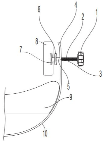
    Figure 1: Parts of the head holder. 1) Handle, 2) Threads, 3) Nut or the hole
in the head holder wall, 4) The wing of the head holder, 5) Shank, 6) The outer
disk, 7) The inner disk, 8) Foam cushion on the fixation axis, 9) Head cushion
below the head, 10) The head holder body.
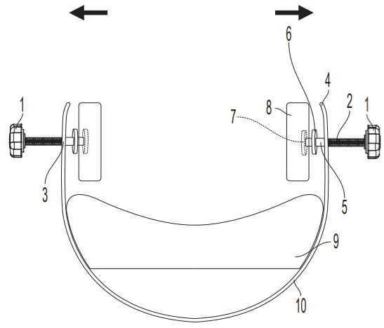
    Figure 2: Parts of the head holder. 1) Handle, 2) Threads, 3) Nut or the
hole in the head holder wall, 4) The wing of the head holder, 5) Shank, 6)
The outer disk, 7) The inner disk, 8) Foam cushion on the fixation axis, 9)
Head cushion below the head, 10) The head holder body. The black arrows
indicates the head cushions can be moved to the sides which allow releasing
of the patient’s head from the immobilization or to allow patients to have more
space to place their heads in the head holder. This depends on a screw like
mechanisms as seen in number 2 of the illustration which is the thread part.
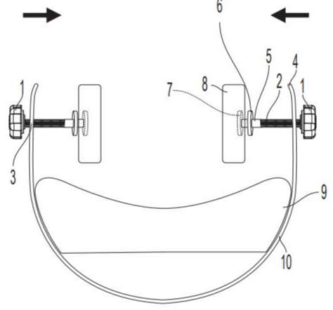
    Figure 3: Parts of the head holder. 1) Handle, 2) Threads, 3) Nut or the hole
in the head holder wall, 4) The wing of the head holder, 5) Shank, 6) The outer
disk, 7) The inner disk, 8) Foam cushion on the fixation axis, 9) Head cushion
below the head, 10) The head holder body. The black arrows indicate the
head cushions can be moved to the center allowing to fix the patient’s head
in the immobilization. This depends on a screw like mechanisms as seen in
number 2 of the illustration which is the thread part.
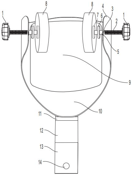
    Figure 4: Parts of the head holder. 1) Handle, 2) Threads, 3) Nut or the hole
in the head holder wall, 4) The wing of the head holder, 5) Shank, 6) The outer
disk, 7) The inner disk, 8) Foam cushion on the fixation axis, 9) Head cushion
below the head, 10) The head holder body, 11) Straight part of the neck, 12)
Diagonal part of the neck, 13) Part inserted in the table, 14) Fixation hole.
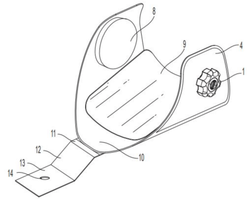
    Figure 5: Parts of the head holder. 1) Handle, 4) The wing of the head holder,
8) Foam cushion on the fixation axis, 9) Head cushion below the head, 10)
The head holder body, 11) Straight part of the neck, 12) Diagonal part of the
neck, 13) Part inserted in the table, 14) Fixation hole.
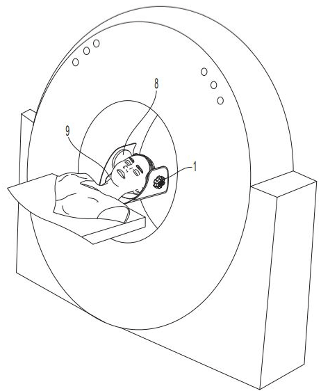
    Figure 6: Parts of the head holder. 1) Handle, 8) Foam cushion on the fixation
axis, 9) Head cushion below the head. This illustrates how to use the head
holder with the patients and be attached to the table of the CT scanner.
Measurements of dimensions
The measurements of the dimensions of the head holder are crucial to build a prototype and to be used with patients. All measurements must be followed except the width of the head holder it can be increased to allow scanning different head sizes and allow more freedom to immobilize any head size (Figure 7 and 8).
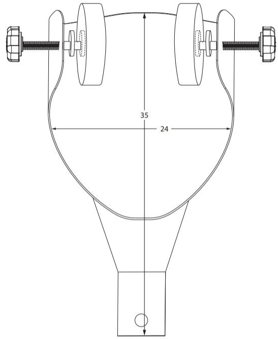
    Figure 7: The measurments of the head holder. The length is 35cm and the
width is 24cm. The width can be increases when manufacturing a prototype
to allow scanning of different head sizes.
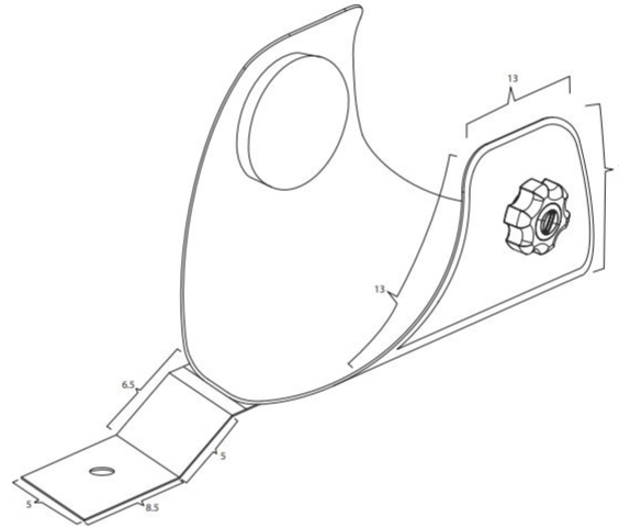
    Figure 8: The measurements of the head holder. The height of the head
holder wing is 13cm, the length is 13cm, the diagonal part of the wing is 13cm,
the straight part of the neck is 1.5cm, the diagonal part of the neck is 5cm,
the length of the part inserted in the table is 8.5cm, and the width of the part
inserted in the table is 5cm.
Discussion
The highest collective dose in diagnostic radiology is made by CT scanners compared to all other imaging modalities [1]. Therefore; repeating a CT scan due to something that can be avoided (i.e. motion artifact by tremors) is desirable. The repeat will increase the radiation dose, contrast media volume, and cost [2]. Sedation can be used with patients who have involuntary movements in case if they do not have any heart problems. More than 31% of the artifact is motion artifacts [3]. The rate of motion artifact varies from 1.9% to 82.7% of the patients and re-exposure rate is 6.4% of the patients [4]. Therefore, this firm head holder design will prevent any motion artifact in patients who suffer severe uncontrolled tremors.
Conclusion
This new design for head holder can fix and immobilize the patients with severe tremor. A prototype should be made then a further study should be conducted to evaluate the success rate for this product and effectiveness. As well, is this head holder can be used in other imaging modalities?
References
- Masjedi H, Zare MH, Keshavarz Siahpoush N, Razavi-Ratki SK, Alavi F, Shabani M. European trends in radiology: Investigating factors affecting the number of examinations and the effective dose. La radiologia medica. 2020; 125: 296-305.
- Doda Khera R, Singh R, Homayounieh F, Stone E, Redel T, Savage CA, et al. Deploying clinical process improvement strategies to reduce motion artifacts and expiratory phase scanning in chest CT. Scientific Reports. 2019; 9: 1-7.
- Veikutis V, Budrys T, Basevicius A, Lukosevicius S, Gleizniene R, Unikas R, et al. Artifacts in computer tomography imaging: how it can really affect diagnostic image quality and confuse clinical diagnosis. Journal of Vibroengineering. 2015; 17: 995-1003.
- Moratin J, Berger M, Rückschloss T, Metzger K, Berger H, Gottsauner M, et al. Head motion during cone-beam computed tomography: Analysis of frequency and influence on image quality. Imaging Science in Dentistry. 2020; 50: 227.