Research Article
Austin J Radiol. 2022; 9(2): 1192.
Renal Segmental Artery Resistive Index as a Non-Invasive Indicator of Functional Deterioration in Patients with Chronic Kidney Disease
Neupane NP1*, Koirala K2, Koirala S3, Lohani B4
1Department of Radiodiagnosis and Imaging, Shahid Gangalal National Heart Centre, Bansbari, Kathmandu, Nepal
2Department of Radiodiagnosis and Imaging, Nepal Medical College Teaching Hospital, Jorpati, Kathmandu
3Nepalese Army Institute of Health Sciences, Syanobharyang, Kathmandu
4Department of Radiodiagnosis and Imaging, Tribhuvan University Teaching Hospital, Maharajgunj, Kathmandu
*Corresponding author: Nirmal Prasad Neupane, Radiologist, Shahid Gangalal National Heart Centre, House No 6/12, Tulasi Marg, Kaushaltar, Bhaktapur, Tel: +9779841914787, Nepal
Received: April 20, 2022; Accepted: May 18, 2022; Published: May 25, 2022
Abstract
Background: Chronic kidney disease refers to the gradual loss of renal function over time and is a significant public health problem worldwide. Doppler ultrasonography can be used as a non-invasive modality to detect renal macroabnormalities and renal vascular status in these patients. The objective of our study was to assess the role of renal segmental artery resistive index in the evaluation of the renal functional status in patients with different grades of chronic kidney disease.
Methods: A hospital-based study using a non-interventional study design was employed in the study. The inclusion criteria were adult patients between 20 to 60 years of age with chronic renal disease but not undergoing renal replacement therapy. Renal Doppler was performed by a single operator on these patients with a curvilinear probe of 3.2 megahertz. Data were entered in a predesigned proforma, and analysis was done using IBM SPSS version 20.
Results: The mean renal segmental artery resistive index in our study was 0.74 (SD=0.08) for the right kidney and 0.75 (SD=0.07) for the left kidney. A positive correlation was observed between the grade of chronic kidney disease and segmental artery resistive index with an r-value of 0.76 and 0.72 for the right kidney and left kidney, respectively.
Conclusion: The renal segmental artery resistive index can be used as a non-invasive indicator for evaluating patients with chronic kidney disease. It correlates with the renal functional status as estimated by the glomerular filtration rate. It can be used as a marker for deterioration of renal function and can also prognosticate the outcome in patients with chronic kidney diseases.
Keywords: Chronic kidney disease; End-stage renal disease; Renal segmental artery doppler; Resistive index
Introduction
Chronic Kidney Disease (CKD) refers to the gradual loss of renal function over time and is a major health problem throughout the globe [1]. It is diagnosed and graded based on laboratory parameters such as urea and creatinine. Its detection and diagnosis in earlier stages is a great challenge that requires invasive modalities such as renal biopsy. Since ultrasound is routinely used for the evaluation of patients with CKD, Doppler ultrasonography can be used as an alternative non-invasive modality to detect renal macroabnormalities and renal vascular status in these patients. The renal segmental artery resistive index is an index of intrarenal arterial resistance and is calculated by the formula: Resistive Index (RI)= (peak systolic velocity – end diastolic velocity)/peak systolic velocity. Previous studies have shown alteration in the resistive index in various renal diseases to be a good indicator of renal functional status [2-5]. This study was conducted to assess the correlation between renal segmental artery resistive index and the functional status of the kidney in patients with different grades of CKD.
Methods
Our study was a hospital-based and non-interventional type. It was a cross-sectional study conducted in a tertiary level hospital, i.e., Tribhuvan University Teaching Hospital for one year. Ethical approval was taken from the Institutional Review Board (IRB), Institute Of Medicine (IOM). The inclusion criteria were adult patients between 20 to 60 years of age with chronic renal disease but not undergoing renal replacement therapy. A total of 138 patients were included in the study. The estimated Glomerular Filtration Rate (eGFR) was calculated using the Cockcroft-Gault (CG) equation and the patients were categorized into different grades of CKD based on the eGFR.
Renal Doppler was performed by a single operator on these patients with a curvilinear probe of 3.2 MHz. Scanning of the bilateral kidneys was performed. For the right kidney, the patient was made to lie in the supine position and the probe was placed in the right lower intercostal space in the mid axillary line. Longitudinal and transverse views were obtained before the Doppler study. For the left kidney, the patient was made to lie either supine or in the right lateral decubitus position. The probe was placed in the left lower intercostal space in the posterior axillary line. For the left kidney, the position of the transducer was more cephalad and posterior than the right kidney. Before the Doppler study, both longitudinal and transverse views were obtained for the left kidney as well.
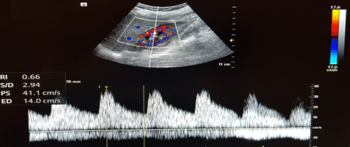
Segmental artery RI was measured in both kidneys at three different sites, i.e., upper, mid, and lower pole, and a mean of the three values was taken (Figure 1). The Doppler angle was kept below 30 degrees in all patients. Data were then entered in a predesigned proforma and analysis was done using SPSS. The ultrasonic findings were correlated with the different grades of CKD using Pearson’s correlation coefficient. The difference in mean of ultrasound parameters between various CKD grades was analysed using one way ANOVA.

    Figure 1: Measurement of the intrarenal resistive index in the mid pole cortex
of the right kidney.
Results
The mean age of the study population was 45.52 years and the age of the patients ranged from 22 to 60 years. Most of the patients were in the age group of 40-50 years (42.8%). The study population comprised of 63% males and 37% females (Table 1). The most common cause of CKD in the study population was hypertension followed by diabetes mellitus.In our study, the majority of the patients were in grade 3 (49.3%) followed by grade 2 (19.6%) (Table 2). Grade 5 comprised the smallest proportion of patients (1.4%).
Parameters
Number (n=138)
(Percentage)
Mean age (SD)
Median age (IQ)
45.52 years
46 years
-
-Age Group
20-30 years
30-40 years
40-50 years
50-60 years
10
27
59
42
(7.2%)
(19.6%)
(42.8%)
(30.4%)Gender
Male
Female
87
51
(63.0%)
(37.0%)
Table 1: Age/gender distribution of the study population.
    GRADE of CKD
    N=138
    Percentage


    Grade    1
    17
    12.3


    Grade    2
    27
    19.6


    Grade    3
    68
    49.3


    Grade    4
    24
    17.4


    Grade    5
    2
    1.4
Table 2: Distribution of study population according to the grade of CKD.
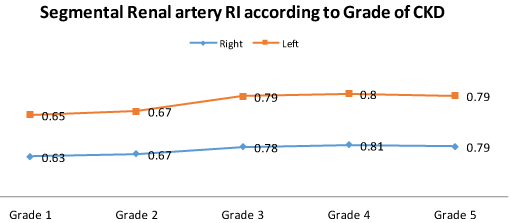
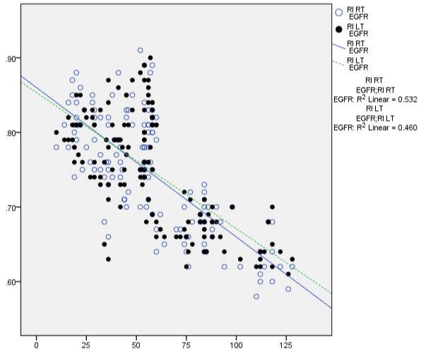
The mean renal segmental artery RI in our study was 0.74 (SD=0.08) for the right kidney and 0.75 (SD=0.07) for the left kidney. The mean segmental artery RI in grade 1 CKD was 0.63 (SD=0.04) for the right kidney and 0.65 (SD=0.03) for the left kidney (Table 3). For grade 2 CKD, the mean segmental artery RI was 0.67 (SD=0.03) for the right and 0.67 (SD=0.02) for the left kidney. Similarly, for grade 3 CKD, the mean segmental artery RI was 0.78 (SD=0.05) for the right and 0.79 (SD=0.06) for the left kidney. For grade 4 CKD, the mean segmental artery RI was 0.81 (SD=0.04) for the right kidney and 0.80 (SD=0.03) for the left kidney. Similarly, for grade 5 CKD, the mean segmental artery RI was 0.79 (SD=0.02) for the right kidney and 0.79 (SD=0.01) for the left kidney. There was a positive correlation between the grade of CKD and segmental artery RI with r-value of 0.76 for the right kidney and 0.72 for the left kidney (Figure 2&3).

    Figure 2: Diagram showing the variation of renal segmental artery RI
according to the grade of CKD.

    Figure 3: Scatter plot diagram showing the correlation between the renal
segmental artery RI and eGFR.
Resistive Index (RI)
Grade 1
Mean(SD)Grade2 Mean(SD)
Grade 3
Mean(SD)Grade 4
Mean(SD)Grade 5
Mean(SD)p-value
RT
LT0.63(0.04)
0.65(0.03)0.67(0.03)
0.67(0.02)0.78(0.05)
0.79(0.06)0.81(0.04)
0.80(0.03)0.79(0.02)
0.79(0.01)<0.001
Table 3: Variation in renal segmental artery RI according to the grade of CKD.
Discussion
In the present study, we have tried to correlate the renal vascular parameter, i.e., renal segmental artery resistive index with the estimated GFR and the grade of the disease. In chronic kidney disease, there is a progressive loss of the nephrons leading to a decrease in the GFR [6,7]. This deterioration in the function of the nephrons leads to the elevation of the serum creatinine and urea with various consequences associated with it such as neuropathy, vasculopathy, and retinopathy which are also included in the spectrum of the chronic kidney diseases. In chronic kidney disease, there is not only loss of nephrons but also of the capillaries. With the progression in the grade of the disease, there is progressive loss of capillaries, and a reduction in the number and luminal area of the intrarenal vessels [3]. There is interstitial fibrosis with reduced caliber of the intrarenal vessels resulting in an increase in the intrarenal vascular resistance [8]. This increase in vascular resistance can be used as an indicator of the progression of the disease [9,10].
The Resistive Index (RI) which reflects the downstream renal artery resistance can be non-invasively measured using the color and spectral Doppler studies. Renal segmental artery RI has been found to correlate with renal vascular resistance, filtration fraction, and effective renal plasma flow in chronic kidney diseases according to previous studies [5,10,11]. Hence renal segmental artery RI can not only classify chronic kidney disease into various stages but can also provide an insight regarding the progression of the disease.
In our study, we found that the renal segmental artery RI increases with the increasing grade of CKD. We found a positive correlation between the grade of CKD and segmental artery RI with an r-value of 0.76 for the right kidney and 0.72 for the left kidney. The segmental artery RI was found to increase with an increase in the grade of CKD from grade 1 to grade 4. However, there was a slight decrease in RI in grade 5 diseases. This was probably because of the small number of patients in grade 5 (only 2 patients) in our study, and we suggest evaluation of a larger group for the same. Overall, there was a positive correlation between the grade of CKD and renal artery RI with an r-value of 0.76 for the right and 0.72 for the left kidney. This finding corresponds to that in the study of Petersen et al., where they found a significant increase in the renal Resistive Index in patients with CKD and found a negative correlation between RI and GFR (r= -0.5, P= 0.02) [10,11]. Previous studies, in addition to the results mentioned above, have also shown that the renal resistive index might predict the rapid versus slow progression of CKD. The limitation of our study was that we were not able to differentiate the slow versus rapid progression of the disease as ours was a cross-sectional study. We hope, this study acting as a pioneer study, will ignite other similar studies in the field so that we have better radiological understanding of the histological vascular alterations in patients with chronic kidney diseases.
Conclusion
The renal segmental artery resistive index can be used as a noninvasive indicator for evaluating patients with chronic kidney disease. It correlates with the renal functional status as estimated by the glomerular filtration rate. It can be used as a marker for deterioration of renal function in patients with chronic kidney diseases.
Acknowledgement
We would like to thank all the participants of the study for their contribution towards this research.
References
- Luca D, Carmine Z. Chronic kidney disease prevalence in the general population: heterogeneity and concerns. Nephrol Dial Transplant. 2015; 31: 331-335.
- Splendiani G, Parolini C, Fortunato L, Sturniolo A, Costanzi S. Resistive index in chronic nephropathies: predictive value of renal outcome. Clinical nephrology. 2002; 57: 45-50.
- Radermacher J, Ellis S, Haller H. Renal resistance index and progression of renal disease. Hypertension. 2002; 39: 699-703.
- Ikee R, Kobayashi S, Hemmi N, Imakiire T, Kikuchi Y, Moriya H, et al. Correlation between the resistive index by Doppler ultrasound and kidney function and histology. American journal of kidney diseases. 2005; 46: 603- 609.
- Sugiura T, Wada A. Resistive index predicts renal prognosis in chronic kidney disease: results of a 4-year follow-up. Clinical and experimental nephrology. 2011; 15: 114-1120.
- Smith HW. The kidney: structure and function in health and disease: Oxford University Press. 1951.
- Ruggenenti P, Schieppati A, Remuzzi G. Progression, remission, regression of chronic renal diseases. The Lancet. 2001; 357: 1601-1608.
- Eardley KS, Kubal C, Zehnder D, Quinkler M, Lepenies J, Savage CO, et al. The role of capillary density, macrophage infiltration and interstitial scarring in the pathogenesis of human chronic kidney disease. Kidney international. 2008; 74: 495-504.
- Kimura N, Kimura H, Takahashi N, Hamada T, Maegawa H, Mori M, et al. Renal resistive index correlates with peritubular capillary loss and arteriosclerosis in biopsy tissues from patients with chronic kidney disease. Clinical and experimental nephrology. 2015; 19: 1114-1119.
- Petersen L, Petersen J, Talleruphuus U, Ladefoged S, Mehlsen J, Jensen H. The pulsatility index and the resistive index in renal arteries.Associations with long-term progression in chronic renal failure. Nephrology Dialysis Transplantation. 1997; 12: 1376-1380.
- Petersen L, Petersen J, Ladefoged S, Mehlsen J, Jensen H. The pulsatility index and the resistive index in renal arteries in patients with hypertension and chronic renal failure. Nephrology Dialysis Transplantation. 1995; 10: 2060-2064.