Clinical Image
Austin J Radiol. 2022; 9(3): 1193.
A Rare Cause of Chronic Chest Pain Syndromes; Scimitar Syndrome Apropos of a CAS
Boukhalit A¹, Nassar H² and Kaukone NR³*
1Department of Radiology, Mohammed V University of Rabat, Central Avicenna Radiology Service, Rabat
2Department of Radiology, Mohammed V University of Rabat, Professor at the Avicenne Central Radiology, Rabat
3Department of Radiology, University Mohammed V of Rabat
*Corresponding author: Kaukone Nyare Raissa Albertine, Department of Radiology, University Mohammed V of Rabat
Received: June 13, 2022; Accepted: July 05, 2022; Published: July 12, 2022
Clinical Image
This is Mr. M, 38 years old History of intermittent chest pain with a type of contraction evolvings in childhood and increased by food.
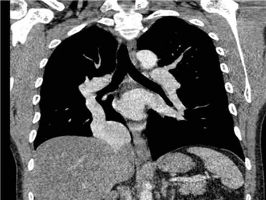
    Coronal thoracic section showing a Pulmonary venous return is ensured by a

large pulmonary vein measuring 15mm in diameter, with a vertical path and

empties directly at the level of the inferior vena cava which is very dilated.

Coronal thoracic section showing a Pulmonary venous return is ensured by a
large pulmonary vein measuring 15mm in diameter, with a vertical path and
empties directly at the level of the inferior vena cava which is very dilated.
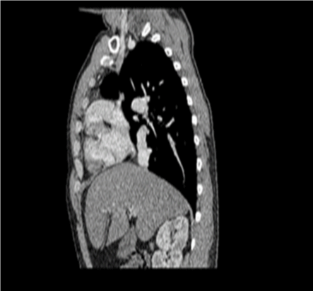
    Axial and sagittal thoracic section showing Pulmonary venous return is

ensured by a large pulmonary vein measuring 15mm in diameter, with a

vertical path and flows directly to the level of the inferior vena cava which is

very dilated

Dilation of the right heart chambers.

Dilatation of the trunk of the pulmonary artery measuring 38 mm and of the

right and left pulmonary arteries and their lobar and permeable dividing

branches after injection of contrast product.
References
- K boubetta, alouikasbi. Congenital pulmonary malformations imaging findings. journal of pediatrics and puericulture. 2004; 17: 370-379.
- N Nakle, S Biscardi, V Lambert, A Sigal-Cinqualbre, R Epaud, F Madhi. Anomalous left coronary artery from pulmonary artery revealed by acute bronchiolitis. Rev Mal Respir. 2012; 29(7): 912-5.
- A Richard, F Godart, G-M Breviere, C Francart, C Foucher, C Rey. abnormal origien of the left coronary artery from the pulmonary artery: a rertrospective study of 36 cases. Arch Mal Coeur Vaiss. 2007; 100(5): 4333-8.
- Ali Dodge-Khatami, Constantine Mavroudis, Carl L Backer. Anomalous origin of the left coronary artery: collective review of surgical therapy. Ann Thorac Surg. 2002; 74(3); 946-55.
- Shaun W Leong, Aidan J Borges, Jessica Henry, Jagdish Butany. Anomalous left coronary artery from the pulmonary artery: case report and review of the literature. Int J Cardiol. 2009; 133(1): 132-4.