Clinical Image
Austin J Radiol. 2022; 9(3): 1194.
Bilateral Ovarian Masses in Krukenberg Tumors
Khouchoua S*, Zahi H, Iraqi Houssaini Z, Jerguigue H, Latib R and Omor Y
Department of Radiology, Ibn Sina University Hospital Center, Morocco
*Corresponding author: Khouchoua S, Department of Radiology, National Institute of Oncology, Ibn Sina University Hospital Center, Avenue Allal El Fassi, 10000, Rabat, Morocco
Received: June 14, 2022; Accepted: July 07, 2022; Published: July 14, 2022
Clinical Image
A 47 years old women with no prior comorbidities, presented with a 3 months history of abdominal distension with insidious pain, weight loss and fatigue.
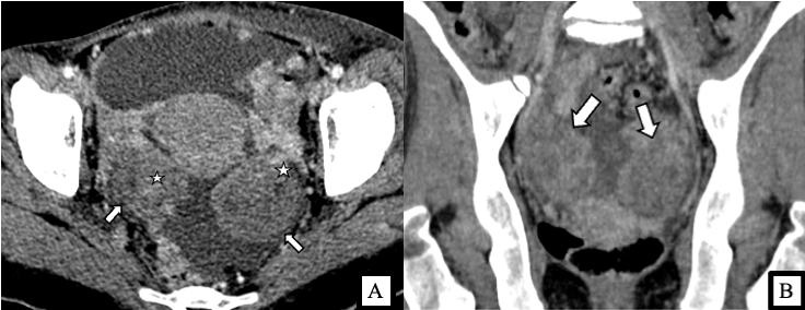
The patient underwent an abdominal computed tomography scan, that showed bilateral ovarian masses, large, well defined, and mixed solid and cystic, with enhancement of the solid component (Figure 1A & 1B).

    Figure 1: Axial (A) and coronal (B) contrast enhanced abdominal CT scan showing enlarged ovaries bilaterally replaced by two well defined masses mixed cystic

and solid (arrows) with enhancement of the solid component (asterisk).
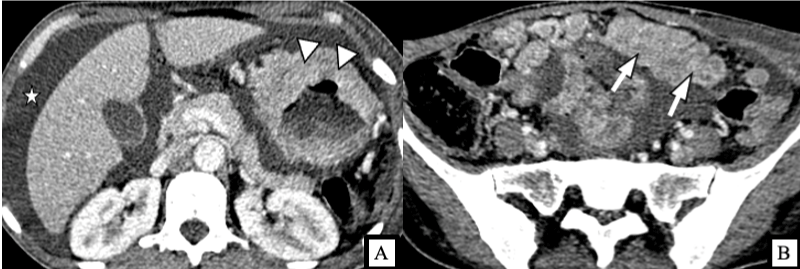
Further examination, revealed gastric wall thickening with large volume ascites (Figure 2A), extensive omental infiltration, and serosal implants (Figure 2B).

    Figure 2A & b): Axial contrast enhanced abdominal CT scan showing irregular wall thickening of the stomach (arrowheads) associated with large volume ascites

(asterisk) and extensive omental thickening with an omental cake appearance (arrows).
Histologic analysis was consistent with stromal infiltration by poorly differentiated adenocarcinoma, immunohistochemical staining showed cytokeratin-7 negativity and cytokeratin-20 positivity favoring a metastatic gastrointestinal carcinoma.
Krukenberg tumors account for 50% of ovarian metastatic masses, with nearly 80% being bilateral [1]. Extraovarian adenocarcinomas with signet ring cells have a predilection for metastasizing to the ovaries. The stomach is the most common primary tumor site followed by the colon, appendix and breast particularly the lobular invasive carcinoma type.
Typically, imaging features of bilateral ovarian masses, solid or mixed solid and cystic with avid enhancement of the solid parts is very suggestive [2]. In fact the bilaterality is a key finding, and in the presence of a gastrointestinal neoplasm, metastatic ovarian Krukenberg tumor should be considered until proven otherwise.
This eponym refers to a pathologic definition; therefore histopathological analysis is necessary to confirm the diagnosis. The prognosis of Krukenberg tumors is very poor and patients may benefit from resection of the primary tumor and ovarian masses when feasible.
Keywords: Krukenberg Tumor; Bilateral Ovarian Mass; Ovarian Metastasis; Signet Ring Cell Adenocarcinoma.
References
- Zulfiqar M, Koen J, Nougaret S, Bolan C, VanBuren W, McGettigan M, Menias C. Krukenberg Tumors: Update on Imaging and Clinical Features. AJR Am J Roentgenol. 2020; 215: 1020-1029.
- Karaosmanoglu AD, Onur MR, Salman MC, Usubutun A, Karcaaltincaba M, Ozmen MN, Akata D. Imaging in secondary tumors of the ovary. Abdom Radiol (NY). 2019; 44: 1493-1505.