
Clinical Image
Austin J Radiol. 2022; 9(3): 1195.
Cytotoxic Lesion of the Corpus Callosum (CLOCCs)
Khouchoua S*, Zahi H, Bourekba I, and Laamrani F-Z
Department of Radiology, Ibn Sina University Hospital Center, Morocco
*Corresponding author: Khouchoua S, Department of Radiology, National Institute of Oncology, Ibn Sina University Hospital Center, Avenue Allal El Fassi, 10000, Rabat, Morocco
Received: June 14, 2022; Accepted: July 07, 2022; Published: July 14, 2022
Clinical Image
A 19 year old male with no past medical history presented to the emergency department with an altered mental status, seizures and fever. Laboratory examination showed no abnormalities, further examination with urine and serum drug screening however, revealed high levels of benzodiazepines. Initial head computed tomography came back normal, therefore an MRI was performed. It featured focal hypersignal lesion of the splenium of the corpus callosum on both T2 and FLAIR (Figure A and B) weighted images with diffusion restriction (Figure D).
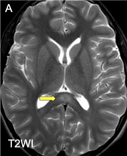
    Figure A: Axial T2weighted image showing a focal hyperintense lesion of the

splenium of the corpus callosum (arrow).
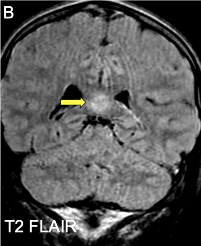
    Figure B: Coronal FLAIR weighted image showing a focal hypersignal of the

splenium of the corpus callosum (arrow).
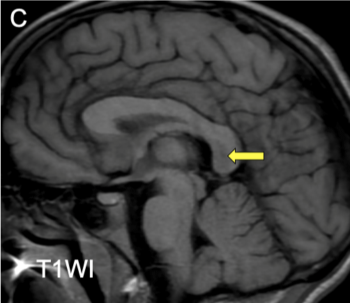
    Figure C: SagittalT1 weighted image showing a focal hyposignal of the

splenium of the corpus callosum (arrow).
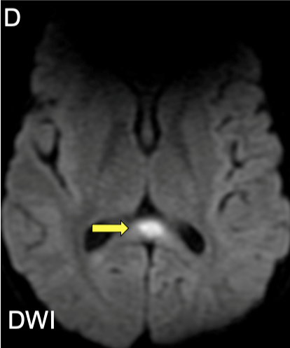
    Figure D: Axial Diffusion weighted image of the forementioned lesion of the

splenium of the corpus callosum featuring diffusion restriction (arrow).
Cytotoxic lesions of the corpus callosum (CLOCCS) are associated to a wide collection of conditions ranging from infection, subarachnoid haemorrhage, malignancy to drug induced and metabolic disorders [1]. It refers to signal changes usually in the splenium, secondary to a stereotyped cascade of cytokines, resulting in massive increase of glutamate thought to be preferentially expressed in the splenium and responsible for a cytotoxic oedema. CLOCCs is to be considered in patients with seizure or confusion along with symptoms related to the underlying causal pathology, although clinical presentation is usually non-specific. Imaging and especially MRI is pivotal to make the diagnosis. In fact, it can only be appreciated on MRI [2] with three possible classical findings: centered lesion of the splenium featuring high intensity signal on T2 (A, arrow) and FLAIR (B, arrow), hyposignal on T1 (C, arrow) and restricted diffusion (D, arrow)with or without extension either to the adjacent white matter laterally or to the anterior corpus callosum. These lesions are known to be reversible after treatment of the underlying pathology with complete healing within a few weeks [2].
Keywords: Splenium; Corpus Callosum; Cytotoxic; Hypersignal.
References