Clinical Image
Austin J Radiol. 2022; 9(3): 1196.
Double Rectum Sign: Anastomotic Leakage
Khouchoua S*, Zahi H, Iraqi Houssaini Z, Jerguigue H, Latib R and Omor Y
Department of Radiology, Ibn Sina University Hospital Center, Morocco
*Corresponding author: Khouchoua S, Department of Radiology, National Institute of Oncology, Ibn Sina University Hospital Center, Avenue Allal El Fassi, 10000, Rabat, Morocco
Received: June 14, 2022; Accepted: July 07, 2022; Published: July 14, 2022
Clinical Image
A 41-year-old patient presents a prolonged ileus with abdominal tendernesss even days after an anterior resection with total excision of the mesorectum and latero terminal colorectal anastomosis for a rectal adenocarcinoma. The laboratory results reveal elevated CRP levels.
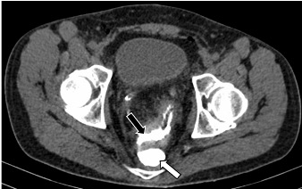
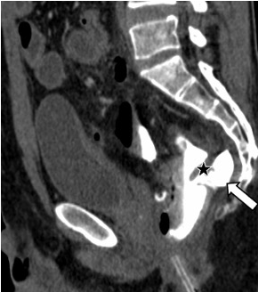
An abdomino-pelvic CT scan with low water-soluble opacification shows a leakage of the contrast media with individualization of a parietal defect next to the colorectal anastomosis (Figure 1), feeding a pre-sacral collection mimicking a rectal lumen in a “double rectum sign” appearance (Figure 2A & B).

    Figure 1: Axial CT image after retrograde opacification showing contrast

media extravasation with a pre sacral collection mimicking the rectal lumen

featuring the double rectum sign (white arrow) adjacent to the true rectum

(black arrow).

    Figure 2: Sagittal CT image demonstrating contrast media leakage (asterisk)

forming a pre sacral collection featuring the double rectum sign (arrow).
Anastomotic leakage is one of the most important complications of colorectal cancer surgery. It refers to a parietal defect involving the anastomosis area, leading to communication between the intra and extra luminal compartments [1]. This complication is suspected in the presence of an infectious syndrome and particularly in the event of significant increase in CRP levels [1].
Imaging is the key to make the correct diagnosis using computed tomography and relies on a rigorous acquisition protocol [2] where rectal opacification with water-soluble fluids plays a major role.
In fact, extravasation of contrast media remains the most reliable sign to make the diagnosis [2].
The “Double rectum sign” appearance is defined by the presence of contrast leakage resulting in a collection that may contain mottled air bubbles or an air fluid level [1]. This collection most often lies at the level of the pre-sacral space, whereby mimicking the rectal lumen and suggesting the diagnosis of an anastomotic leak [2].
It can be associated with extensive fat infiltration or even intra peritoneal extension with multiple collections responsible for suppuration and peritonitis.
Keywords: Anastomotic Leakage; Double Rectum Sign;Colorectal Cancer.
References
- Weinstein S, Osei-Bonsu S, Aslam R, Yee J. Multidetector CT of the postoperative colon: review of normal appearances and common complications. Radiographics. 2013; 33: 515-32.
- Tamini N, Cassini D, Giani A, Angrisani M, Famularo S, Oldani M, Montuori M, Baldazzi G, Gianotti L. Computed tomography in suspected anastomotic leakage after colorectal surgery: evaluating mortality rates after false-negative imaging. Eur J Trauma Emerg Surg. 2020; 46: 1049-1053.