Case Report
Austin J Radiol. 2022; 9(3): 1197.
Exophthalmos it’s not always a Cellulitis: Orbital Lymphoma Case Report
Khouchoua S*, Zahi H, Iraqi Houssaini Z, Jerguigue H, Latib R and Omor Y
Department of Radiology, Ibn Sina University Hospital Center, Morocco
*Corresponding author: Khouchoua S, Department of Radiology, National Institute of Oncology, Ibn Sina University Hospital Center, Avenue Allal El Fassi, 10000, Rabat, Morocco
Received: June 14, 2022; Accepted: July 07, 2022; Published: July 14, 2022
Abstract
Orbital lymphoma is a rare cause of primary Non Hodgkin lymphomasand represents a major cause of orbital malignancies, usually arising from extraocular muscles, eyelid, and soft tissue adnexa. Overall it presents as an extraconal tumor featuring a broad spectrum of clinical presentation making the diagnosis sometimes challenging. We report the case of a 65 year old male with no prior medical history presenting with a recent ongoing proptosis. Radiological findings were consistent with an extraconal lesion involving the lateral orbital wall responsible for mass effect on the globe and optic nerve, for which histopathological results showed features of a small lymphocytic lymphoma.
Keywords: Orbital Lymphoma; Exophthalmos; Cellulitis; CT
Case Presentation
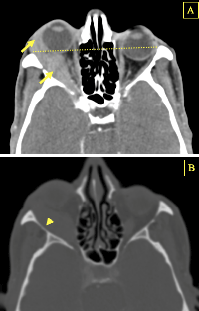
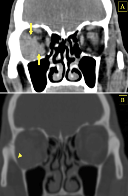
We report the case a 65 year old male with no past medical history presenting to the emergency department for right orbital pain and blurred vision for 15 days with a worsening proptosis for one month prior, associated with recent weight loss and episodes of fever. Patient denied any trauma history. Physical examinations showed a visual acuity of the right eye of 6/10 no evidence of chemosis, swelling or inflammation. Laboratory analysis found normal white blood cell count, and moderately elevated C reactive protein at 25mg/l. The rest of physical examination and laboratory study were unremarkable. A computed tomography of the orbits was performed showing a right orbital mass of soft tissue density enhancing homogeneously after contrast media injection, involving the lateral wall, bulging medially with mass effect on the globe and optic nerve responsible for moderate exophthalmos, associated with infiltration of the lateral rectus muscle and extension to the pre septal soft tissue. Further examination showed no evidence of bone destruction adjacent to the mass and no intra cranial extension or through the optic canal, superior or inferior orbital fissures. Although clinical presentation with recent onset of orbital pain, vision impairment and episodes of fever initially was suspicious of orbital cellulitis, both laboratory studies and imaging findings clinched the diagnosis of an orbital tumor which characteristics were highly suggestive of a lymphoma. Patient then underwent a biopsy of the mass, histopathological analysis showed a diffuse small lymphocytic infiltration and immunochemical study concluded to a marginal zone B cell lymphoma.
Discussion
Orbital lymphomas and especially Non-Hodgkin’s type represent less than 2% of all lymphomas [1]. Among the broad spectrum of histological subtypes, the vast majority of cases are reported to be extra nodal marginal zone lymphomas [2]. Nonetheless orbital lymphoma remains the most common primary orbital neoplasm in the adult population with increasing incidence over the last decade [3]. It arises in most cases from mucosa associated lymphoid tissue, and the potential role of a previous Chlamydia psittaci exposure is thought to play a major role in the pathogenesis among other infective processes [3].
Orbital lymphoma is a disease of the elderly between 50 and 70 years of age. Gender predilection depends on the histological subtype. In fact, extranodal marginal zone B-cell lymphoma and follicular lymphoma tend to have a female predilection, whereas diffuse large B-cell lymphomas show an even gender distribution [4].
It can either involve the conjunctiva, eyelid, orbital walls or even the lacrimal gland [5].
Patients usually present with a palpable mass causing orbital swelling [4]. Other symptoms like diplopia, extraocular movement limitation, or decreased visual acuity can be found, but proptosis remains the hallmark symptom. Vision impairment can be caused by optic nerve infiltration or compression through mass effect. Sometimes clinical presentation can be misleading especially when symptoms overlap with those of orbital cellulitis like in our case [6].
Radiological analysis on both Computed Tomography (CT) and/or Magnetic Resonance Imaging (MRI) plays a major role in the characterization of orbital masses and more commonly in the exploration of an exophthalmos.
Exophthalmos or proptosis as a key clinical and radiological feature refers to the anterior displacement of the globe. The reference line on cross sectional imaging is the inter zygomatic line. The lower limit of normal distance from this line to the posterior aspect of the globe is around 6mm below which we can conclude to a proptosis.
Regarding imaging characteristics, on Computed Tomography (CT) orbital lymphoma appears as a soft tissue mass involving any part of the orbit with a predilection for the upper outer quadrant usually close to the lacrimal gland.
It is usually isodense or moderately hyperdense compared to extra ocular muscles on non-enhanced CT examination, with discrete to mild and homogeneous enhancement after contrast media administration.
Mass effect is also a main feature, on the extra ocular muscles, globe and optic nerve hence the protrusion and exophthalmos on one the hand and decrease in vision acuity on the other hand [7].
When important mass effect is present, especially on the surrounding muscles, it can be challenging to delineate the origin of the mass, MRI is therefore useful as it offers better contrast resolution. Nevertheless, in most cases we can easily determine the origin of the mass with respect to the extra ocular muscles on CT, particularly helpful in making the correct diagnosis of lymphoma and to differentiate it from other orbital masses especially idiopathic orbital inflammation that most commonly involves the extra ocular muscles and is considered the most challenging differential diagnosis. In addition, with regards to clinical presentation, especially if symptoms are misleading like in our case, orbital cellulitis can also be among the differentials [5]. Close analysis of imaging feature scan help clinch the diagnosis, in the absence of extensive pre or post septal fat stranding, orbital abscess or contiguous sinusitis.
In addition, imaging plays a pivotal role in the diagnostic approach in differentiating lymphoma from other orbital neoplasms with rare globe or optic nerve invasion and bone destruction. Some authors even suggest that within orbital lymphoma’s subtypes, radiological features like globe indentation is significantly more likely to be encountered in aggressive subtypes, but no notable difference was found otherwise, on both CT and MRI [2] among orbital lymphomas. Pathological confirmation remains mandatory for diagnostic purposes and appropriate treatment management.
Radiotherapy is considered the gold standard for localized extra nodal primary orbital lymphoma with no evidence of systemic involvement [8].
Otherwise, for patients with systematic or metastatic spread, no specific treatment guideline exists and surgical resection, radiotherapy, and chemotherapy can be used; rituximab being the monoclonal antibody most commonly utilized for the treatment of CD 20 positive small lymphocytic lymphoma [9]. Recent identification of Chlamydia psittaci lead to the use of antibiotic therapy with some reported cases of decrease in tumor size and even remission [3].
Therefore, optimal treatment strategy must be discussed by a multidisciplinary team depending on clinical, laboratory and radiological data.

    Figure 1: Axial contrast enhanced CT images on both soft tissue (A) and

bone windows (B) showing a right orbital mass involving the lateral wall, of

soft tissue density enhancing homogeneously after contrast media injection,

bulging medially with mass effect on the globe and optic nerve responsible

for moderate exophthalmos above the inter zygomatic line, associated with

infiltration of the lateral rectus muscle and extension to the pre septal soft

tissue (arrows)with no evidence of bone erosion or destruction (arrow head).

    Figure 2: Coronal contrast enhanced CT images on both soft tissue (A) and

bone windows (B) showing a right orbital mass involving the lateral wall,

of soft tissue density with mass effect on the optic nerve and the lateral

and inferior rectus muscles (arrows)with no evidence of bone erosion or

destruction (arrow head).
Our patient had no evidence of systematic dissemination and radiotherapy was therefore the treatment of choice.
Localized orbital non-Hodgkin’s lymphoma’s prognosis depend on the histological subtype. In fact, mucosa associated lymphoid tissue (MALT) as one type of marginal zone lymphoma (MSL) seems to have good prognosis with a 5 year relapse free rate around 65%, and low mortality rates not exceeding 5% [4,10].
Conclusion
Non-Hodgkin’s orbital lymphoma is a major cause of proptosis with an overall good prognosis.
However the broad variety of clinical presentation can sometimes be misleading.
Imaging plays a pivotal role in lesion characterization of orbital masses, having an appropriate diagnostic approach with good knowledge of orbital lymphoma imaging features appears to be crucial for early diagnosis, prompt and appropriate management.
Conflict of Interest
The authors declare no conflicts of interest.
Funding Acknowledgement
This research received no funding.
References
- Ahmed S, Shahid RK, Sison CP, Fuchs A, Mehrotra B. Orbital Lymphomas: A Clinicopathologic Study of a Rare Disease. The American Journal of the Medical Sciences. 2006; 331: 79-83.
- Juniat V, Cameron CA, Roelofs K, Bajic N, Patel S, Slattery J, et al. Radiological analysis of orbital lymphoma histological subtypes. Orbit. 2022; 1-9.
- Moslehi R, Devesa SS, Schairer C, Fraumeni JF. Rapidly increasing incidence of ocular non-hodgkin lymphoma. Journal of the National Cancer Institute. 2006; 142: 895-896.
- Olsen TG, Heegaard S. Orbital lymphoma. Surv Ophthalmol. 2019; 64: 45- 66.
- Coupland SE, White VA, Rootman J, Damato B, Finger PT. A TNM-based clinical staging system of ocular adnexal lymphomas. Archives of pathology & laboratory medicine. 2009; 133: 1262-7.
- Chaurasiya BD, Agrawal G, Chaudhary S, Shah S, Pradhan A, Lavaju P. Orbital Lymphoma Masquerading as Orbital Cellulitis. Case Reports in Ophthalmological Medicine. 2021; 2021: 1-5.
- Serodio JF, Jonet M, Trinidade M, Coutinho I, Favas C. A Rare Case of Bilateral Proptosis. European Journal of Case Reports in Internal Medicine. 2018; 5: 1.
- Stafford SL, Kozelsky TF, Garrity JA, Kurtin PJ, Leavitt JA, Martenson JA, et al. Orbital lymphoma: radiotherapy outcome and complications. Radiotherapy and oncology: journal of the European Society for Therapeutic Radiology and Oncology. 2001; 59: 139-144.
- Esmaeli B, McLaughlin P, Pro B, Samaniego F, Gayed I, Hagemeister F, et al. Prospective trial of targeted radioimmunotherapy with Y-90 ibritumomab tiuxetan (Zevalin) for front-line treatment of early-stage extranodal indolent ocular adnexal lymphoma. Annals of oncology: official journal of the European Society for Medical Oncology. 2009; 20: 709-714.
- Ollila TA, Olszewski AJ. Extranodal Diffuse Large B Cell Lymphoma: Molecular Features, Prognosis, and Risk of Central Nervous System Recurrence. Current Treatment Options in Oncology. 2018; 19: 1-20.