Case Report
Austin J Radiol. 2022; 9(4): 1200.
Imaging of Omphalopagus: A Case Report
Mribat M*, El Haddad S, Tantaoui S, Chat L and Allali N
Department of Radiology, Pediatric Teaching Hospital, Mohammed V University, Morocco
*Corresponding author: Mribat M, Department of Radiology, Pediatric Teaching Hospital, Mohammed V University, Rabat, Morocco
Received: June 30, 2022; Accepted: July 25, 2022; Published: August 01, 2022
Abstract
Omphalopagus is a form of conjoined twins with relatively good prognosis. In this rare congenital anomaly, the union of the twins starts at the lower part of the thorax and extends to the umbilical region. We relate the case of conjoined omphalopagus twins lately diagnosed by ultrasonography and foetal MRI at 28-weeks gestation. Case report: We received a 28 years-old primigravid nulliparous patient at 28-weeks gestation, diagnosed with omphalopagus conjoined twins by ultrasonography. We then proceeded with foetal MRI to further assess the health of the twins. Conclusion: Although an early diagnosis by ultrasound can be auspicious, the use of foetal MRI late in a conjoined twins pregnancy can be a great tool to help the surgical team plan their therapeutic strategy.
Keywords: Conjoined twins, MRI, Omphalopagus
Introduction
Conjoined twins are a rare developmental anomaly of monozygotic monoamniotic twin gestation. They result from the incomplete division of the embryonic disc beyond thirteen days of gestation [1]. They’re classified according to their main site of fusion. Early prenatal diagnosis is based essentially on ultrasound and is mandatory for follow up and management.
Observation
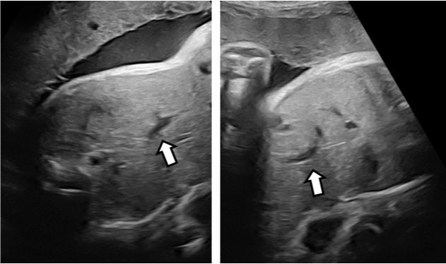
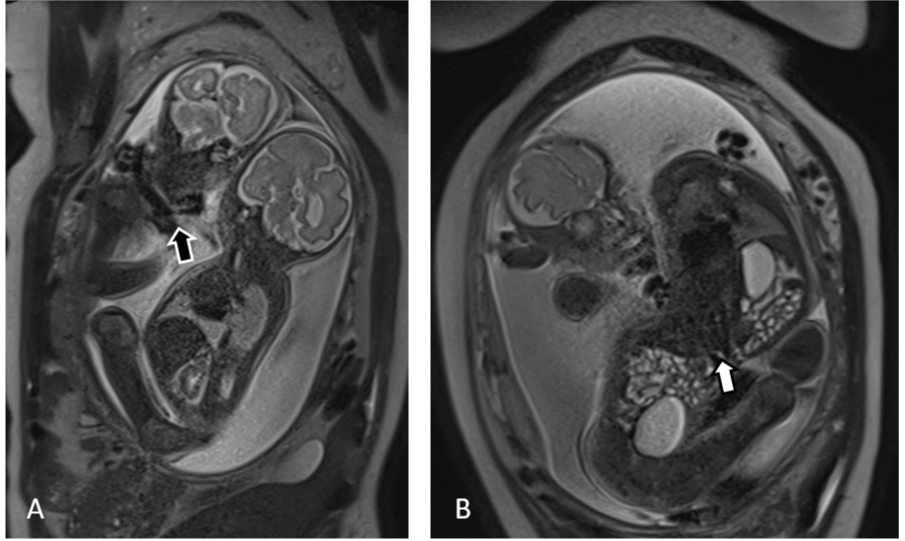
We relate the case of a 28 years old patient, primigravid nulliparous, with no history of consanguinity or other known risk factor. Her gynaecologist for a routine ultrasonography examination at 28-weeks gestation referred her. Sonography revealed monochorionic-monoamniotic twins who were attached by their abdomen, starting from the lower thorax to their umbilic, sharing their diaphragm and their liver (Figure 1). Each foetus had 4 limbs, his own central and peripheral nervous system, his gastro-intestinal tract, his gallbladder and portal sinus (Figure 2) as well as his urinary tract and cardio-respiratory system. We proceeded with foetal MRI (Figures 3 and 4) to confirm the ultrasonography results and to make a more exhaustive assessment of the twins’ health. After ruling out other malformations, we made the diagnosis of omphalopagus.

    Figure 1: Ultrasound images showing the point of union (A) of the twins and

their hearts (B) arrows.

Figure 2: Ultrasound images (A,B) showing the portal sinus of each twin (arrows).

Figure 3: Foetal MRI : T2 HASTE axial images (A-C) showing the shared liver (A), shared distal intestinal tract (B) and shared umbilical chord (C).

Figure 4: Foetal MRI: T2 HASTE coronal image (A) showing nuchal chord (black arrow). T2 HASTE coronal image (B) showing the fusion of the twins at their livers.
Discussion
Although Siamese twins are frequent in some areas of South-East Asia and Africa, with a frequency reaching 1:14 000 to 1:25 000, it is a pretty rare condition elsewhere in the world, with an incidence approaching 1:50 000 to 1:200 000. Females are more involved with a ratio ranging between 1.6 and 3 [2–5].
The exact etiopathogenesis of conjoined twins is still indefinite. No chromosomic anomaly was detected. Nor race, heredity or consanguinity seems to be involved [6]. In the literature, two opposing theories try to give an explanation to this conundrum. The classic one states that there would be an incomplete division of the embryo after 14 days of gestation. While the other one indicates that the siamese result of partial fusion of two separate embryonic discs [7]. Many classifications are used in the literature to describe the different types of Siamese twins (Figure 5). From their attachment site, we can distinguish the thoracopagus (thorax), omphalopagus (abdomen), pyopagus (sacrum), ischiopagus (pelvis), craniopagus (skull), cephalopagus (face) and rachipagus (spine) [8,9].

Figure 5: Classification of the different Forms of conjoined twins.
The most reported forms of conjoined twins are the thoracopagus, the omphalopagus and the thoraco-omphalopagus, representing 70% of siamese twins described in the literature [10].
Conjoined twins are usually discovered during the first trimester of gestation, between the 10th and 13th week of gestation. Although, diagnosis can be assumed as early as the 9th week of gestation. Ultrasonography contributes to the diagnosis by demonstrating the fused organs so we can classify the type of conjoined twins. And it’s also helpful with the assessment of additional malformations that may deteriorate even further the prognosis.
In our case, we diagnosed the omphalopagus on ultrasonography by revealing the fusion from the lower part of the thorax to the umbilical region. In this form of Siamese, the liver is fused 80% of the time. The pericardium can also be shared but not the heart. Ultrasonography must analyse the liver parenchyma, the portal system and the inferior vena cava of each foetus. If a twin has an abnormal or absent portal system, his survival after surgery is compromised [11].
Moreover, ultrasound examination must analyse the intra and extra-hepatic biliary tree as well as the pancreas and the number of gallbladders. Usually, the stomach and proximal intestinal tract are separate [12]. The colon divides distally and each twin has his own rectum and a set of upper and lower extremities. There’s no pelvic or urinary union.
Other techniques like CT and foetal MR can be used later to evaluate the prognosis. With its excellent anatomical definition, we used foetal MR to better analyse the liver parenchyma, its vascular supply, the biliary system and the gastro-intestinal tract. At 28 weeks of gestation, the foetal MR helped us confirm the ultrasonography results and rule out other malformations that could have modified therapeutic management of the siamese.
Unfortunately, our patient came to us late during her second trimester of pregnancy. At that stage, we recommended a monthly ultrasonographic monitoring while we wait for the surgical team plan.
Survival of conjoined twins is usually very low and results vary with the site and complexity of the conjunction. In their study, Romero et al. reported a 39% of mortality at birth [13].
Conclusion
Conjoined twins are a rare malformation. Prognosis depends on the extent and the location of the conjunction. Prenatal imaging with early ultrasonography plays a major role in the diagnosis of this kind of anomaly and helps with the screening of other malformations. Foetal MR is more useful later during the pregnancy to assess the prognosis. The challenge for the radiologist is to make the most precise diagnosis in order for the surgical team to adapt their management strategy.
References
- Konan Blé R, Séni K, Adjoussou S, Quenum G, Akaffou E, Koné M. Jumeaux conjoints craniopages difficultés de prise en charge en milieu africain. Gynécologie Obs Fertil. 2008; 36: 56–59.
- Hanson JW. Incidence of Conjoined Twinning. 1975; 306: 1257.
- Edmonds LD, Layde PM. Conjoined Twins in the United States, 1970–1977. Teratology. 1982; 25: 301–310.
- Diaz JH, Furman EB. Perioperative management of conjoined twins. Anesthesiology. 1987; 67: 965–973.
- Wilson RL, Cetrulo CL, Shaub MS. The prepartum diagnosis of conjoined twins by the use of diagnostic ultrasound. Am J Obstet Gynecol. 1976; 15: 737.
- Guèye M, Guèye SMK, Guèye MDN, Diouf AA, Niang MM, Diallo M, et al. Accouchement de jumeaux conjoints de découverte fortuite au cours du travail au CHU de Dakar. Pan Afr Med J. 2012; 12: 102.
- Benirschke K, Kaufmann P. Pathology of the Human Placenta. Springer New York; 2000.
- Mathew RP, Francis S, Basti RS, Suresh HB, Rajarathnam A, Cunha PD, et al. Conjoined twins – role of imaging and recent advances. J Ultrason. 2017; 17: 259–266.
- Pierro A, Kiely EM, Spitz L. Classification and clinical evaluation. Semin Pediatr Surg. 2015; 24: 207–211.
- Sharma UK, Dangol A, Chawla CD, Shrestha D. Antenatal detection of conjoined twin. J Nepal Med Assoc. 2007; 46: 133–135.
- O’Neill JA, Holcomb GW, Schnaufer L, Templeton JM, Bishop HC, Ross AJ, et al. Surgical experience with thirteen conjoined twins. Ann Surg. 1988; 208: 299.
- Spitz L. Conjoined twins. Br J Surg. 1996; 83: 1028–1030.
- Bieber FR. Prenatal diagnosis of congenital anomalies. Appleton and Lange, East Norwalk. Teratology. 2022; 22: 354–355.