Research Article
Austin J Radiol. 2022; 9(4): 1201.
Research on Anatomy of the Pulp Chamber Floor of the Maxillary Second Permanent Molar in CBCT
Xiuyou W¹, Tiwari SK², Jinhong J³, Yao X², Ling Y² and Li P²*
¹Department of Pediatric Dentistry, Affiliated Xiangya Stomatological Hospital & Xiangya School of Stomatology, Central South University, China
²Department of Cariology and Endodontics, Affiliated West China Hospital of Stomatology of Sichuan University, China
³Department of Oral and Maxillofacial Surgery, Affiliated the Chinese People’s Liberation Army 921 Hospital of Joint Logistics Support Force, China
*Corresponding author: Peng Li, Department of Cariology and Endodontics, West China Hospital of Stomatology of Sichuan University, No.14, Section 3, Renmin South Road, Chengdu, China
Received: July 18, 2022; Accepted: August 16, 2022; Published: August 23, 2022
Abstract
Objective: The objective of this study was to examine the pulp chamber floor anatomy of the maxillary second permanent molar in Chinese individuals by using Cone-Beam Computed Tomography (CBCT). These data may facilitate endodontic treatment success.
Methodology: A total of 2505 CBCT images of maxillary second permanent molars were studied to evaluate the shape of the pulp chamber floor, the types of developmental root fusion lines (DRFLs), the number of canal orifices and their possible relationships.
Results: Three pulp chamber floor shapes were identified: triangular (50.3%), rhomboid (34.3%) and oval (15.4%). The shape of the DRFLs and the number of canal orifices on the pulp chamber floor were variable. The frequency of non branching DRFLs was 63.8%, followed by branching DRFLs (33.6%). The I-shaped DRFL group had more variations in the number and location of canal orifices.
Conclusion: The anatomy of the pulp chamber floor of the maxillary second permanent molar in Chinese individuals is diverse. Knowledge of variations in pulp chamber shape, types of DRFLs and canal orifice number of the maxillary second permanent molar can help reduce the risk of missing root canals in endodontic treatment.
Keywords: Maxillary second molar; CBCT; Pulp chamber floor; DRFL; Canal orifice
Introduction
A satisfactory outcome of Root Canal Treatment (RCT) is achieved after complete cleaning, shaping and obturating the root canal system. To accomplish this goal, a systematic understanding of pulp chamber floor anatomy and the root canal system is necessary [1]. The maxillary second permanent molar is located in the posterior corner of the dental arch. Variations in tooth morphology, a complicated root canal system, deposition of secondary dentin and limited operation space increase the difficulty of RCT. Anatomical variations in terms of morphology and number of roots and canals have been reported [2-22]. However, analysis of the pulp chamber floor anatomy of the maxillary second permanent molar is inadequate. Since the pulp chamber floor is the first impression of the root canal system, the study of the anatomy of the pulp chamber floor can help clinicians precisely locate the canal orifices and reduce the risk of missing root canals.
There are two approaches to studying the morphology of teeth, namely, invasive and noninvasive procedures. The number of samples associated with invasive procedures is limited, and teeth cannot be reused for further research. Conventional two-dimensional radiography is an important noninvasive tool for investigating root canal morphology and pulp chamber position. However, it is difficult to identify the number of canal orifices, the location of canal orifices on the pulp chamber floor and variations in the morphology of the root canal system with two-dimensional radiographs [2-6]. Currently, noninvasive and three-dimensional imaging approaches for studying tooth anatomy are widely performed. Cone-Beam Computed Tomography (CBCT) is one of the most favored methods for studying tooth morphology because the images produced by CBCT are as accurate as those obtained by clearing and modified staining methods [6], and they are produced with relatively lower doses of radiation and shorter working times [23].
The anatomy of the pulp chamber floor in multirooted teeth is complex. The canal orifices present as a dark mark with a funnel shape on the pulp chamber floor. The canal orifices are interconnected by grooves on the pulp chamber floor. These grooves were first described as “subpulpar grooves” and “depressions of the pulp chamber floor” [24]. Krasner and Rankow described subpulpar grooves as “Developmental Root Fusion Lines” (DRFLs), which are darker than the pulp chamber floor color. DRFLs may present as deep or shallow grooves, and the law of color change in the pulp floor is a better method for identifying DRFLs and orifices located on the pulp floor [25]. DRFLs are observed during direct observation of the pulp chamber floor and can be regarded as important trace marks for orifice location.
Although studies have noted the anatomical variations in the pulp chamber floor in the maxillary second permanent molar [2- 22,26], the correlation between DRFLs and the orifice location on the pulp chamber floor has not been studied. The aim of this study was to investigate the shape of the pulp chamber floor of the maxillary second permanent molar with CBCT and evaluate the types of DRFLs, the number of canal orifices and their possible relationships.
Materials and Methodology
A total of 2505 CBCT images of the maxillary second permanent molar from 1789 patients were obtained from the medical image center of ××Hospital of Stomatology, ××University, ××, P.R. China. The 3D Accuitomo CBCT machine (MCT-1[EX-2F], J. Morita Manufacturing Corp, Kyoto, Japan) used for the study produces images of 14-bit grayscale, 0.125 mm voxel size and 1 mm thickness. The teeth included had a closed apex, no restoration, and a history of RCT or periapical surgery. Examination of coronal, axial and sagittal sections of images was performed by moving the toolbar from the crown to the apex of the root to identify the shape of the pulp chamber, shape of the DRFLs, and site and number of root canal orifices on the pulp chamber floor. One-volume Data Viewer software (J. Morita Manufacturing Corp) was used by two endodontists to inspect the images.
Results
The age of the patients included in the study was between 15 and 84 years old, with an average age of 39.79 years. Among the 1789 patients, 871 (48.7%) were male, and 918 (51.3%) were female.
Shape of the Pulp Chamber Floor of the Maxillary Second Permanent Molar
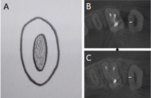
The shape of the pulp chamber floor was studied at the level of the Cemento Enamel Junction (CEJ). Three shapes of the pulp chamber floor, namely, triangular, rhomboid and oval, were identified (Figure 1). The presence of a triangular-shaped pulp chamber floor was found at a higher frequency (50.3%) than those for rhomboid-shaped (34.3%) or oval-shaped (15.4%) floors (Table 1).
    Shape of pulp chamber

    Triangular

    Rhomboid

    Oval


    N(%)

    1261(50.3)

    857(34.3)

    387(15.4)
Table 1: Distribution of shapes of pulp chamber floor in maxillary second permanent molar.

    Figure 1: (A) Illustrations show the three shapes of pulp chamber in maxillary second permanent molar. (B) Cone-beam computed tomography images show the

three shapes of pulp chamber in maxillary second permanent molar. The arrows indicate the examined tooth.
Types of Drfls in the Maxillary Second Permanent Molar
1. The dark line on the pulp chamber floor was described as the DRFL. In this study, DRFLs were found in 2441 cases. According to their appearance, the DRFLs were divided into three types and six subtypNo DRFL: a tooth without a DRFL because there was only one canal orifice in the chamber floor (Figure 2).

    Figure 2: Illustration of maxillary second permanent molar showed the presence of the pulp chamber floor with no DRFL(A). CBCT axial planes of tooth with one

orifice on pulp chamber floor (B). CBCT axial planes which are 3mm apical to pulp chamber floor (C). Arrows indicate the orifice.
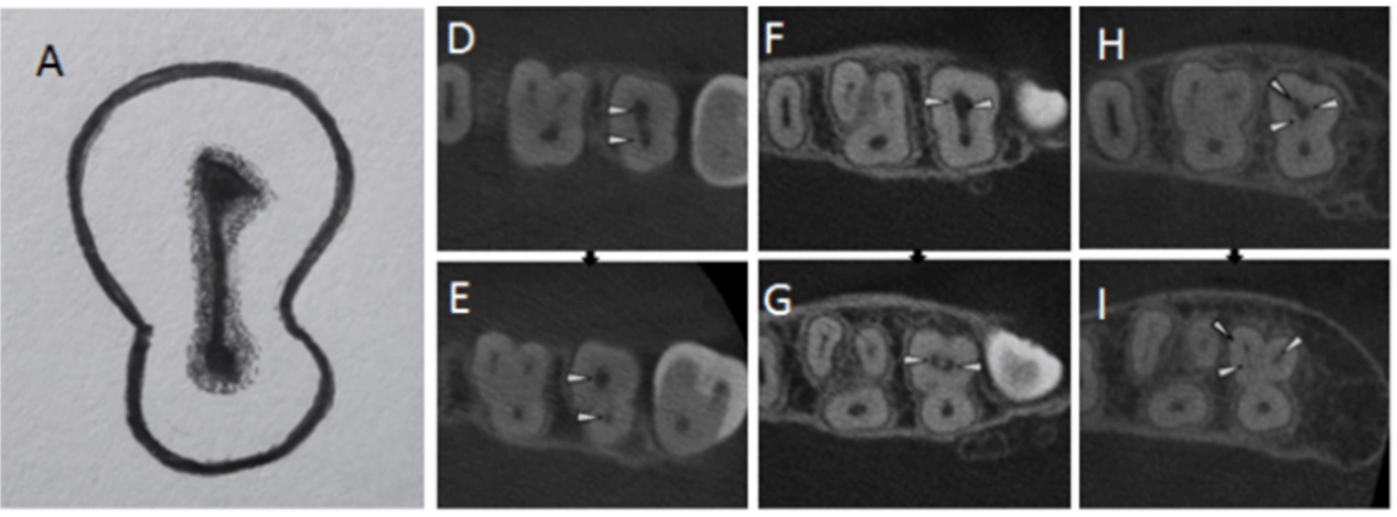
2. Nonbranching DRFL: A line starting from the palatal canal orifice end in the buccal orifice without producing any distributions. The line is identified as an I-shaped DRFL when it is a straight line from the palatal canal orifice to the buccal canal orifice (Figure 3A). If the line has some degree of angulation while passing forward, it is identified as a 7-shaped DRFL (Figure 4A) or inverted U-shaped DRFL (Figure 4F).

    Figure 3: Illustration of maxillary second permanent molar showed the presence of I-shaped DRFL on pulp chamber floor (A). CBCT axial planes of tooth with

four sub-types of I-shaped DRFL on pulp chamber floor (D, F, H). CBCT axial planes which are 3mm apical to pulp chamber floor for each of the sub-type (E,G, I).

Arrows indicate the orifice and canal.

    Figure 4: Illustration of maxillary second permanent molar shows the presence of 7-shaped DRFL (A) and inverted U-shaped DRFL (F) on pulp chamber floor.

CBCT axial planes of tooth with two sub-types of 7-shaped DRFL (B, D) and invertedU-shaped DRFL (G) on pulp chamber floor. CBCT axial planes which are 3mm

apical to pulp chamber floor for each of the sub-type (C, E, H). Arrows indicate the orifice and canal.
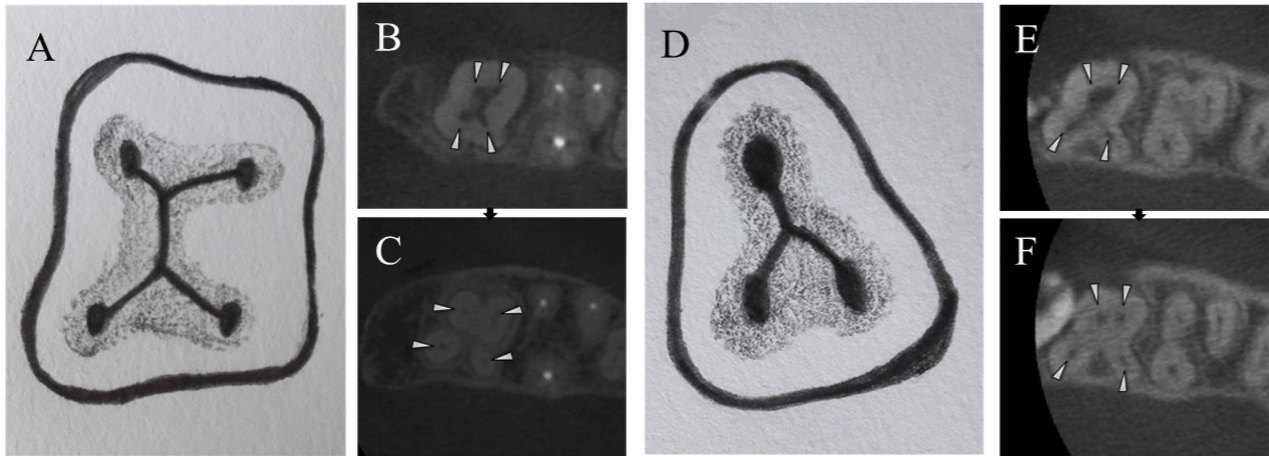
3. Branching DRFL: a line proceeding toward the buccal direction that splits into branches and passes forward to join the respective canal orifices, similar to a Y shape (Figure 5). While in the teeth with two palatal canal orifices, the two proceeding lines from each palatal canal orifice join together and divide or may proceed as a single line in the buccal direction to end in respective buccal canal orifices as an X-shaped DRFL (Figure 6A) and an inverted Y-shaped DRFL (Figure 6D).

    Figure 5: Illustration of maxillary second permanent molar shows the presence of Y-shaped branching DRFL on pulp chamber floor (A,D,G). CBCT axial planes of

tooth with two sub-types of Y-shaped DRFL on pulp chamber floor (B,E,H,J). CBCT axial planes which are 3mm apical to pulp chamber floor for each of the subtype

(C,F,I,K). Arrows indicate the orifice and canal.

    Figure 6: Illustration of maxillary second permanent molar shows the presence of X-shaped branching DRFL on pulp chamber floor (A). CBCT axial planes of tooth

with X-shaped (B) and inverted Y-shaped (E) on pulp chamber floor. CBCT axial planes which are 3mm apical to pulp chamber floor for each of the sub-type (C,F).

CBCT axial planes of tooth showing the bulge on cemento-enamel junction at palatal side indicate the presence of second palatal canal orifice on pulp chamber

floor (B,E). Arrows indicate the orifice and canal.
The distributions of the different types of DRFLs on the pulp chamber floor in the maxillary second permanent molar are shown in Table 2. DRFLs were not found in 64 cases, which had only one canal orifice in the center of the chamber floor. Most teeth had a nonbranching DRFL (63.8%, 1598/2505), while 33.6% of all the cases showed a branching DRFL on the pulp chamber floor. For the nonbranching DRFLs, the 7 shape was found in the highest frequency (68.1%), followed by the I shape (31.0%) and inverted U shape (0.9%). Nearly 97% of the cases of branching DRFLs were Y-shaped DRFLs. The chamber floor shape of all the cases with X-shaped and inverted Y-shaped DRFLs were classified as rhomboid. The 7-shaped and Y-shaped DRFLs were commonly present in cases of triangularshaped pulp chambers. The chamber floors of more than 50% of the cases with I-shaped DRFLs and without DRFLs were identified as oval shaped.
Table 2: Distribution of different types of DRFL on Pulp chamber floor in maxillary second permanent molar.
Distribution of Root Canal Orifices in Cases of Different Types of Drfls
No DRFL
A single orifice was present in the center of the pulp chamber floor. For this case, there was only one canal orifice (Figure 2).
Nonbranching DRFL
I-shaped DRFL: The buccal end of the DRFL had variable numbers of canal orifice(s): either one buccal orifice (Figure 3D and 3E), two buccal orifices (Figure 3F and 3G) or three buccal orifices (Figure 3H and 3I). The canal orifices at the buccal end were close to each other. The palatal end had only one palatal canal orifice in this type. I-shaped DRFLs with a single buccal orifice had the highest prevalence (53.1%), followed by two buccal orifices (35%) and three buccal orifices (11.9%) (Table 3).
Table 3: The distribution of Root Canal orifice in subtypes of DRFL.
7-shaped DRFL: This type was subdivided into two categories noted by capital letters. A. The Mesiobuccal (MB), Distobuccal (DB) and Palatal (P) canal orifices were connected by deep grooves (Figure 4B and 4C), which were identified in 61.3% of the cases (Table 3). B. The 7-shaped DRFL consisted of the second Mesiobuccal (MB2) canal orifice as well as the MB, DB and P canal orifices (Figure 4D and 4E). According to our data, 38.7% of the cases showed three buccal orifices on the pulp chamber floor.
Inverted U-shaped DRFL
The second palatal orifice was present in all cases of this type (Figure 4F). A tooth with the second palatal (P2) canal orifice had a palatal bulge on the external surface of the tooth (Figure 4G and 4H). Therefore, all the cases with inverted U-shaped DRFLs had two palatal orifices. Most cases (92.9%) showed two buccal orifices. Only one case presented three buccal orifices.
Branching DRFL
A. Y-shaped DRFL: This DRFL passes from the palatal canal orifice and divides into mesial and distal branches. All the cases in this group had only one palatal canal orifice (Table 3). However, the ends of the buccal branches had a variable number of canal orifices. This group was subdivided into three categories noted by capital lettThe MB, DB and P canal orifices were connected by Y-shaped superficial (Figure 5B) or deep grooves (Figure 5E). A total of 86.9% of the cases in the Y-shaped DRFL group had two buccal orifices.
B. Four distinct canal orifices (MB, MB2, DB and P) were present on the pulp chamber floor. The location of the MB2 canal orifice was variable: it was located close to the MB canal orifice (Figure 5H and 5I) or at the junction of the line joining the MB and DB canal orifices (Figure 5J and 5K). A total of 13.0% of the cases in the Y-shaped DRFL group had three buccal orifices.
C. Five canal orifices [MB, MB2, DB, second distobuccal (DB2) and P] were present on the pulp chamber floor. However, only one case in the Y-shaped DRFL group had four buccal orifices.
X-Shaped and Inverted Y-Shaped Drfls
In the X-shaped DRFL type, two palatal canal orifices (P and P2) were present on the pulp chamber floor. DRFLs stretch from both palatal canal orifices and join together before reaching the buccal side and connect the MB and DB canal orifices (Figure 6B and 6C). In some of the cases, the MB and DB canal orifices were located close to each other or were connected by deep grooves, and this type of DRFL formed an inverted Y-shaped pattern (Figure 6E). Most of the cases with the X-shaped and inverted Y-shaped DRFLs showed two buccal orifices and two palatal orifices. However, only one case in the X-shaped DRFL group had three buccal orifices, and one case in the inverted Y-shaped DRFL group had one buccal orifice (Table 3).
Most of the teeth had three canal orifices (62.0%, 1552 of 2505), followed by four canal orifices (24.9%, 623 of 2505) (Table 4). Among the teeth with four canal orifices, the incidences of the MB2 and P2 canal orifices were 94.1% (586/623) and 5.9% (37/623), respectively. The two canal orifice cases were all observed in the I-shaped DRFL group. In addition, there were three cases with five canal orifices on the pulp chamber floor. One of them had two root canal orifices in the mesial aspect (MB and MB2 canal orifices), two distal aspects (DB and DB2 canal orifices) and one palatal canal orifice. The other two cases had one MB canal orifice, two distal canal orifices (DB and DB2) and two palatal canal orifices (P and P2).
1
2
3
4
5
N(%)
64(2.5)
263(10.5)
1552(62.0)
623(24.9)
3(0.1)
Table 4: Number of Root Canal orifices in Maxillary second permanent molar.
Discussion
The objective of root canal therapy, eradication of bacteria from the root canal system and restoration of tooth function, can be achieved through cleaning, shaping and obturating the root canal system. Therefore, access preparation is the first and most important aspect of RCT. The ideal access provides an unimpeded passageway for an instrument to reach the apical foramen through the root canal of the tooth, which can facilitate adequate cleaning and shaping and minimize the risk of instrument separation in the root canal during the procedure. The shape of the pulp chamber determines the shape of the access opening. It has been reported that the shape of the pulp chamber is 1, rhomboid shaped in teeth with four canal orifices, 2, triangular shaped in teeth with three canal orifices and 3, oval shaped in teeth with buccal and palatal canal orifices [27]. The findings of this study are consistent with our data in terms of the shape of pulp chambers. However, the CBCT images of the maxillary second molars in our study show the variable number of canal orifices on the floor of all three shapes of pulp chambers.
The correlation between DRFLs and canal orifice location on the pulp chamber floor of the maxillary second permanent molar has not been reported before. The data in this study show a close relationship between DRFLs and the location of canal orifices. This relationship highlights the need to explore canal orifices on the pulp chamber floor along the DRFL during RCT. When a complete DRFL cannot be found during treatment, a canal may have been missed.
The number of canal orifices on the pulp chamber floor is variable in maxillary second permanent molars. According to our study, three canal orifices were present in 62% (1552/2505 cases) of the cases. Almost all three canal orifice cases showed two buccal orifices and one palatal orifice, except 1 case with one buccal orifice and two palatal orifices. This result is consistent with a previous study [26]. The most frequently encountered extra canal orifice was the MB2 canal orifice. MB2 was present in 23.4% (586/2505 cases) of the cases in our study, and this frequency is within the range of previously published papers [10-22]. These prevalence differences among different studies could be due to the type of study approach (in vivo vs. in vitro), the technique of studying root canal morphology (CBCT examination, modified canal staining and clearing technique, spiral computed tomography, and plain and contrast medium-enhanced digital radiography), or an ethnic difference.
The presence of a P2 canal orifice is a rare phenomenon, but its existence cannot be overlooked. The frequency of a P2 root canal in the maxillary second molar has been reported, and its prevalence ranges from 0.4% to 2.08% [7-9,28]. In this study, the P2 canal was only found in inverted-U-, X-and inverted-Y- shaped DRFL types and accounted for 1.6% (39/2505 cases) of all the cases. The pulp chamber of the maxillary second molar with two palatal canal orifices is wider on the palatal side, and the access opening must be rectangular in shape [29]. The preparation of conventional oval- or triangular-shaped access openings is a possible reason for missing the second palatal canal orifice during RCT. Therefore, knowledge about variation in pulp floor morphology is valuable for successful RCT. The presence of a third palatal canal has been reported once by Pasternak et al. [4]. In our study, no case with three palatal canals was found, but this possibility cannot be ignored during endodontic practice.
The preoperative analysis of the root canal system was performed by studying the intraoral radiographs. The radiograph produces a two-dimensional image, and the image of the tooth structure is frequently overlapped by adjacent tooth structures, increasing the possibility of misinterpretation of the image and misleading diagnosis. Studies have shown that more canal orifices can be located with magnification aids than with the naked eye [30-32], and CBCT images are free of limitations posed by conventional radiographs [33]. The CBCT scanner used for this study provides high-resolution images that are most suitable for the study of root canal orifice sites and morphology of the root canal system. Thus, the adoption of newer technologies and a better understanding of the anatomy of the pulp floor can ensure the success of RCT.
Conclusions
The maxillary second molar has variations in the shape of the pulp chamber floor, types of DRFLs, and location and number of canal orifices on the DRFLs. Our data can help locate the canal orifices of the maxillary second permanent molar and achieve good endodontic treatment results.
Acknowledgement
The authors deny any conflicts of interest.
References
- Setzer FC, Boyer KR, Jeppson JR, Karabucak B, Kim S. Long-term prognosis of endodontically treated teeth: a retrospective analysis of preoperative factors in molars. Journal of endodontics. 2011; 37: 21-25.
- Holderrieth S, Gernhardt CR. Maxillary molars with morphologic variations of the palatal root canals: a report of four cases. J Endod. 2009; 35: 1060-5.
- Jafarzadeh H, Javidi M, Zarei M. Endodontic retreatment of a maxillary second molar with three separate buccal roots. Australian endodontic journal: the journal of the Australian Society of Endodontology Inc. 2006; 32: 129-132.
- Júnior BP, Teixeira CDS, Silva RG, Vansan LP, Neto MDS. Treatment of a second maxillary molar with six canals. Australian endodontic journal : the journal of the Australian Society of Endodontology Inc. 2007; 33: 42-45.
- Qun L, Longing N, Qing Y, Yuan L, Jun W, Qingyue D. A case of asymmetric maxillary second permanent molar with double palatal roots. Quintessence Int. 2009; 40: 275-6.
- Neelakantan P, Subbarao C, Subbarao CV. Comparative evaluation of modified canal staining and clearing technique, cone-beam computed tomography, peripheral quantitative computed tomography, spiral computed tomography, and plain and contrast medium-enhanced digital radiography in studying root canal morphology. Journal of endodontics. 2010; 36: 1547- 1551.
- Libfeld H, Rotstein I. Incidence of four-rootedmaxillary second permanent molars: literature review and radiographicsurvey of 1,200 teeth. J Endod. 1989; 15: 129-31.
- Peikoff MD, Christie WH, Fogel HM. The maxillary second permanent molar: variations in the number of roots and canals. Int Endod J. 1996; 29: 365-9.
- Kim Y, Lee SJ, Woo J. Morphology of maxillary first and second molars analyzed by cone-beam computed tomography in a korean population: variations in the number of roots and canals and the incidence of fusion. J Endod. 2012; 38: 1063-8.
- Vertucci FJ. Root canal anatomy of the human permanent teeth. Oral surgery, oral medicine, and oral pathology. 1984; 58: 589-599.
- Gilles J, Reader A. An SEM investigation of the mesiolingual canal in human maxillary first and second molars. Oral surgery, oral medicine, and oral pathology. 1990; 70: 638-643.
- Pécora JD, Woelfel JB, Neto MDS, Issa EP. Morphologic study of the maxillary molars. Part II: Internal anatomy. Brazilian dental journal. 1992; 3: 53-7.
- Eskoz N, Weine FS. Canal configuration of the mesiobuccal root of the maxillary second molar. Journal of endodontics. 1995; 21: 38-42.
- Alavi AM, Opasanon A, Ng Y, Gulabivala K. Root and canal morphology of Thai maxillary molars. International endodontic journal. 2002; 35: 478-485.
- Ng YL, Aung TH, Alavi A, Gulabivala K. Root and canal morphology of Burmese maxillary molars. International endodontic journal. 2001; 34: 620- 630.
- Zhang R, Yang H, Yu X, Wang H, Hu T, Dummer PMH. Use of CBCT to identify the morphology of maxillary permanent molar teeth in a Chinese subpopulation. International endodontic journal. 2011; 44: 162-169.
- Zhao YM, Xu X, Sun J, Qiang YL, Qi QG. In vitro study of the secondary mesiobuccal canal of the maxillary second permanent molar. Hua Xi Kou Qiang Yi Xue Za Zhi. 2009; 27: 509-11, 515.
- Lee JH, Kim KD, Lee JK, Park W, Jeong JS, Lee Y, et al. Mesiobuccal root canal anatomy of Korean maxillary first and second molars by cone-beam computed tomography. Oral Surg Oral Med Oral Pathol Oral Radiol Endod. 2011; 111: 785-91.
- Stropko JJ. Canal morphology of maxillary molars: clinical observations of canal configurations. Journal of endodontics. 1999; 25: 446-450.
- Jing YN, Ye X, Liu DG, Zhang ZY, Ma XC. [Cone-beam computed tomography was used for study of root and canal morphology of maxillary first and second molars]. Beijing da xue xue bao. Yi xue ban = Journal of Peking University. Health sciences. 2014; 46: 958-62.
- Silva EJ, Nejaim Y, Silva AI, Haiter-Neto F, Zaia AA, Cohenca N. Evaluation of root canal configuration of maxillary molars in a Brazilian population using cone-beam computed tomographic imaging: an in vivo study. J Endod. 2014; 40: 173-6.
- Kim Y, Lee SJ, Woo J. Morphology of maxillary first and second molars analyzed by cone-beam computed tomography in a korean population: variations in the number of roots and canals and the incidence of fusion. J Endod. 2012; 38: 1063-8.
- Patel S, Dawood A, Ford TP, Whaites E. The potential applications of cone beam computed tomography in the management of endodontic problems. International endodontic journal. 2007; 40: 818-830.
- Vigouroux SAA, Bosaans SAT. Anatomy of the pulp chamber floor of the permanent maxillary first molar. Journal of endodontics. 1978; 4: 214-219.
- Krasner P, Rankow HJ. Anatomy of the pulp-chamber floor. Journal of endodontics. 2004; 30: 5-16.
- Zhao Yu-mei, Qiang Yan-li, Fang Dong-dong, Qiu Hong-liang, Liu Dao-feng, Zong Min, et al. Anatomy and application of pulp-chamber floors in maxillary second permanent molars. Shandong Medical Journal. 2009-42.
- Vertucci FJ, Haddix JE. Tooth morphology and access cavity preparation. In: Cohen S, Hargreaves KM, eds. Pathways of the Pulp.10th ed. St. Louis: Mosby Elsevier. 2011: 136.
- Caliskan MK, Pehlivan Y, Sepetçioglu F, Türkün M, Tuncer SS. Root canal morphology of human permanent teeth in a Turkish population. Journal of endodontics. 1995; 21: 200-204.
- Aggarwal V, Singla M, Logani A, Shah N. Endodontic management of a maxillary first molar with two palatal canals with the aid of spiral computed tomography: a case report. J Endod. 2009; 35: 137-9.
- Buhrley LJ, Barrows MJ, BeGole EA, Wenckus CS. Effect of magnification on locating the MB2 canal in maxillarymolars. J Endod. 2002; 28: 324-7.
- Schwarze T, Baethge C, Stecher T, Geurtsen W. Identification of second canals in the mesiobuccal root of maxillary first and secondmolars using magnifying loupes or an operating microscope. Aust Endod J. 2002; 28: 57- 60.
- Alaçam T, Tinaz AC, Genç O, Kayaoglu G. Second mesiobuccal canal detection in maxillary first molars using microscopy and ultrasonics. Australian endodontic journal : the journal of the Australian Society of Endodontology Inc. 2008; 34: 106-109.
- Patel S, Dawood A, Mannocci F, Wilson R, Ford TP. Detection of periapical bone defects in human jaws using cone beam computed tomography and intraoral radiography. International endodontic journal. 2009; 42: 507-515.